

Reticular Pseudodrusen: Interreader Agreement of Evaluation on OCT Imaging in Age-Related Macular Degeneration
- PMID: 37292179
- PMCID: PMC10244688
- DOI: 10.1016/j.xops.2023.100325
Reticular Pseudodrusen: Interreader Agreement of Evaluation on OCT Imaging in Age-Related Macular Degeneration
Abstract
Purpose: To determine the interreader agreement for reticular pseudodrusen (RPD) assessment on combined infrared reflectance (IR) and OCT imaging in the early stages of age-related macular degeneration across a range of different criteria to define their presence.
Design: Interreader agreement study.
Participants: Twelve readers from 6 reading centers.
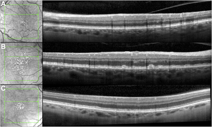
Methods: All readers evaluated 100 eyes from individuals with bilateral large drusen for the following: (1) the presence of RPD across a range of different criteria and (2) the number of Stage 2 or 3 RPD lesions (from 0 to ≥ 5 lesions) on an entire OCT volume scan and on a selected OCT B-scan. Supportive information was available from the corresponding IR image.
Main outcome measures: Interreader agreement, as assessed by Gwet's first-order agreement coefficient (AC1).
Results: When evaluating an entire OCT volume scan, there was substantial interreader agreement for the presence of any RPD, any or ≥ 5 Stage 2 or 3 lesions, and ≥ 5 definite lesions on en face IR images corresponding to Stage 2 or 3 lesions (AC1 = 0.60-0.72). On selected OCT B-scans, there was also moderate-to-substantial agreement for the presence of any RPD, any or ≥ 5 Stage 2 or 3 lesions (AC1 = 0.58-0.65) and increasing levels of agreement with increasing RPD stage (AC1 = 0.08, 0.56, 0.78, and 0.99 for the presence of any Stage 1, 2, 3, and 4 lesions, respectively). There was substantial agreement regarding the number of Stage 2 or 3 lesions on an entire OCT volume scan (AC1 = 0.68), but only fair agreement for this evaluation on selected B-scans (AC1 = 0.30).
Conclusions: There was generally substantial or near-substantial-but not near-perfect-agreement for assessing the presence of RPD on entire OCT volume scans or selected B-scans across a range of differing RPD criteria. These findings underscore how interreader variability would likely contribute to the variability of findings related to the clinical associations of RPD. The low levels of agreement for assessing RPD number on OCT B-scans underscore the likely challenges of quantifying RPD extent with manual grading.
Financial disclosures: Proprietary or commercial disclosure may be found after the references.
Keywords: Age-related macular degeneration; Drusen; OCT; Reticular pseudodrusen; Subretinal drusenoid deposits.
© 2023 by the American Academy of Ophthalmology.
Figures

References
-
- Wu Z., Fletcher E.L., Kumar H., et al. Reticular pseudodrusen: a critical phenotype in age-related macular degeneration. Prog Retin Eye Res. 2022;88 - PubMed
-
- Spaide R.F., Ooto S., Curcio C.A. Subretinal drusenoid deposits AKA pseudodrusen. Surv Ophthalmol. 2018;63:782–815. - PubMed
-
- Sivaprasad S., Bird A., Nitiahpapand R., et al. Perspectives on reticular pseudodrusen in age-related macular degeneration. Surv Ophthalmol. 2016;61:521–537. - PubMed
LinkOut - more resources
Full Text Sources
Research Materials
