. 2023 May 19:13:13.
doi: 10.25259/JCIS_29_2023. eCollection 2023.

Intraperitoneal anatomy with the aid of pathologic fluid and gas: An imaging pictorial review

Affiliations

Intraperitoneal anatomy with the aid of pathologic fluid and gas: An imaging pictorial review

Matthew Montanarella et al. J Clin Imaging Sci. .

Abstract

The peritoneum is a large serosal membrane enveloping the abdomen and pelvic organs and forming the peritoneal cavity. This complex relationship forms many named abdominopelvic spaces, which are frequently involved in infectious, inflammatory, neoplastic, and traumatic pathologies. The knowledge of this anatomy is essential to the radiologist to localize and describe the extent of the disease accurately. This manuscript provides a comprehensive pictorial review of the peritoneal anatomy to describe pathologic fluid and gas.

Keywords: Pathologic fluid; Pathologic gas; Peritoneal anatomy; Peritoneal spaces; Peritoneum.


Conflict of interest statement

There are no conflicts of interest.

Figures

Figure 1:
Figure 1:
Illustrations (a and b) and a normal computed tomography abdomen and pelvis coronal reformatted image (c) reviewing the anatomy of the peritoneum.
Figure 2:
Figure 2:
A 32-year-old woman with bowel perforation. Contiguous computed tomography axial images of the abdomen with IV contrast (a and b) demonstrating an abscess within the right subphrenic space (star). The white arrow indicates the falciform ligament, which separates the right subphrenic space into the right and left sides.
Figure 3:
Figure 3:
A 45-year-old man with bowel perforation. (a) Abdominal computed tomography (CT) scout image and (b) CT abdomen axial image on lung window demonstrating large volume pneumoperitoneum within the right subphrenic space (star). The falciform ligament (arrow) is noted, which separates the right subphrenic space into the right and left subphrenic spaces.
Figure 4:
Figure 4:
A 53-year-old man with liver cirrhosis. Computed tomography abdomen axial image with IV contrast in a patient with ascites demonstrating the subdivisions of the left subphrenic space. The star indicates the immediate subphrenic space. The white and red arrows indicate the left subhepatic and perisplenic spaces.
Figure 5:
Figure 5:
A 29-year-old woman with tubo-ovarian abscess. Computed tomography abdomen axial image with IV contrast demonstrating an abscess within the immediate left subphrenic space (star). The white arrow indicates the gastrophrenic ligament.
Figure 6:
Figure 6:
A 66-year-old male with hepatocellular carcinoma. (a and b) contiguous computed tomography axial images of the abdomen with IV contrast demonstrating a ruptured left hepatic lobe hepatocellular carcinoma (white arrow) and hematoma within the left subhepatic space (star). The red arrow indicates active contrast extravasation.
Figure 7:
Figure 7:
A 30-year-old man with blunt abdominal trauma. (a and b) Contiguous computed tomography axial images of the abdomen with IV contrast demonstrating a splenic laceration (white arrow) and a hematoma in the perisplenic space (star) in a patient with blunt abdominal trauma. The red arrow indicates the phrenicocolic ligament.
Figure 8:
Figure 8:
A 34-year-old man with bowel perforation. Computed tomography abdomen axial image with IV contrast demonstrating an abscess within the anterior right subhepatic space (star). The white arrow indicates the gastrohepatic ligament.
Figure 9:
Figure 9:
A 37-year-old woman with sepsis and intraperitoneal infection. (a) Computed tomography (CT) abdomen axial image demonstrating abscess (white arrow) within Morrison’s pouch. A 40-year-old man status post gunshot wound to the abdomen. (b) CT abdomen axial image demonstrating blood products (red arrow) within Morrison’s pouch.
Figure 10:
Figure 10:
A 62-year-old woman with stage four cholangiocarcinoma. Computed tomography abdomen and pelvis axial image (a), coronal (b), and sagittal (c) reformatted images demonstrating a metastatic peritoneal deposit in the posterior right subhepatic space (white arrow). A peritoneal mass is seen between the liver parenchyma and the right abdominal wall (red arrow). A large metastatic mass is seen centered within the pelvis (star).
Figure 11:
Figure 11:
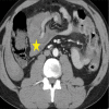
A 52-year-old-man with pancreatitis. Computed tomography abdomen axial image with IV contrast demonstrating a small fluid collection in the superior recess of the lesser sac (white arrow).
Figure 12:
Figure 12:
A 49-year-old man with a history of pancreatitis. Computed tomography abdomen axial image with IV contrast demonstrating a pancreatic pseudocyst (star) in the inferior recess of the lesser sac.
Figure 13:
Figure 13:
A 63-year-old man with hemorrhagic pancreatitis. Computed tomography abdomen axial image with IV contrast demonstrating a pseudocyst in the splenic recess of the lesser sac (star).
Figure 14:
Figure 14:
A 53-year-old woman with disseminated TB and peritoneal ascites. (a through c) Contiguous computed tomography axial images of the abdomen with IV contrast demonstrating omental thickening (white arrows).
Figure 15:
Figure 15:
A 60-year-old male with stage four bladder carcinoma. Axial (a) and coronal reformatted image (b) demonstrating nodular thickening at the bladder dome (red arrow), omental metastasis (white arrows). Of note is a large metastatic deposit within the small bowel mesentery (star) and innumerable enlarged mesenteric lymph nodes.
Figure 16:
Figure 16:
A 41-year-old man with blunt abdominal trauma. (a and b) Contiguous computed tomography axial images of the abdomen with IV contrast (a and b) demonstrating hematoma in the transverse mesocolon (stars).
Figure 17:
Figure 17:
A 44-year-old man with blunt abdominal trauma. Computed tomography abdomen axial image with IV contrast demonstrating a hematoma within the small bowel mesentery (star).
Figure 18:
Figure 18:
A 47-year-old man with bowel perforation. Computed tomography abdomen axial image with IV contrast demonstrating contrast extravasation into the right and left pericolic gutters (white arrows).
Figure 19:
Figure 19:
A 43-year-old man with blunt abdominal trauma. computed tomography abdomen axial image with IV contrast demonstrating a hematoma within the intersigmoid recess (star).
Figure 20:
Figure 20:
Illustration demonstrating the ligaments and peritoneal spaces of the pelvis.
Figure 21:
Figure 21:
A 57-year-old woman with large volume peritoneal ascites. (a-d) Contiguous computed tomography axial images of the abdomen with IV contrast in a patient with large volume ascites, which well delineates the pelvic ligaments. Arrow in image (a) indicates the broad ligament. Arrow in image (b) indicates the round ligament. Arrows in image (c) indicate the paired medial umbilical ligaments. Arrow in image (d) indicates the median umbilical fold.

Similar articles

Cited by

References

    1. Wasnik AP, Maturen KE, Kaza RK, Al-Hawary MM, Francis IR. Primary and secondary disease of the peritoneum and mesentery: Review of anatomy and imaging features. Abdom Imaging. 2015;40:626–42. doi: 10.1007/s00261-014-0232-8. - DOI - PubMed
    1. Levy AD, Arnáiz J, Shaw JC, Sobin LH. From the archives of the AFIP: Primary peritoneal tumors: Imaging features with pathologic correlation. Radiographics. 2008;28:583–607. doi: 10.1148/rg.282075175. - DOI - PubMed
    1. Levy AD, Shaw JC, Sobin LH. Secondary tumors and tumorlike lesions of the peritoneal cavity: Imaging features with pathologic correlation. Radiographics. 2009;29:347–73. doi: 10.1148/rg.292085189. - DOI - PubMed
    1. Meyers MA, Oliphant M, Berne AS, Feldberg MA. The peritoneal ligaments and mesenteries: Pathways of intraabdominal spread of disease. Radiology. 1987;163:593–604. doi: 10.1148/radiology.163.3.3575702. - DOI - PubMed
    1. Feldman GB, Knapp RC. Lymphatic drainage of the peritoneal cavity and its significance in ovarian cancer. Am J Obstet Gynecol. 1974;119:991–4. doi: 10.1016/0002-9378(74)90021-0. - DOI - PubMed

LinkOut - more resources