

This is a preprint.
Needle guidance with Doppler-tracked polarization-sensitive optical coherence tomography
- PMID: 37292463
- PMCID: PMC10246070
Needle guidance with Doppler-tracked polarization-sensitive optical coherence tomography
Update in
-
Needle guidance with Doppler-tracked polarization-sensitive optical coherence tomography.J Biomed Opt. 2023 Oct;28(10):102910. doi: 10.1117/1.JBO.28.10.102910. Epub 2023 Oct 4. J Biomed Opt. 2023. PMID: 37799938 Free PMC article.
Abstract
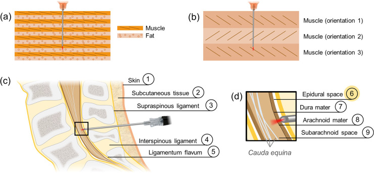
We demonstrate that a simple, unscanned polarization-sensitive optical coherence tomography needle probe can be used to perform layer identification in biological tissues. Broadband light from a laser centered at 1310 nm was sent through a fiber that was embedded into a needle, and analysis of the polarization state of the returning light after interference coupled with Doppler-based tracking allowed the calculation of phase retardation and optic axis orientation at each needle location. Proof-of-concept phase retardation mapping was shown in Atlantic salmon tissue, while axis orientation mapping was demonstrated in white shrimp tissue. The needle probe was then tested on the ex vivo porcine spine, where mock epidural procedures were performed. Our imaging results demonstrate that unscanned, Doppler-tracked polarization-sensitive optical coherence tomography imaging successfully identified the skin, subcutaneous tissue, and ligament layers, before successfully reaching the target of the epidural space. The addition of polarization-sensitive imaging into the bore of a needle probe therefore allows layer identification at deeper locations in the tissue.
Conflict of interest statement
Conflict of interest The authors have no potential conflicts of interest to disclose.
Figures


References
-
- Finlayson Louise, Barnard Isla RM, McMillan Lewis, Ibbotson Sally H, Brown C Tom A, Eadie Ewan, and Wood Kenneth. Depth penetration of light into skin as a function of wavelength from 200 to 1000 nm. Photochemistry and Photobiology, 98(4):974–981, 2022. - PubMed
-
- Petterson Ingeborg E Iping, Day John CC, Fullwood Leanne M, Gardner Benjamin, and Stone Nick. Characterisation of a fibre optic Raman probe within a hypodermic needle. Analytical and Bioanalytical Chemistry, 407:8311–8320, 2015. - PubMed
-
- Alix James JP, Plesia Maria, Hool Sarah A, Coldicott Ian, Kendall Catherine A, DBE Pamela J Shaw, Mead Richard J, and Day John C. Fiber optic Raman spectroscopy for the evaluation of disease state in Duchenne muscular dystrophy: An assessment using the mdx model and human muscle. Muscle & Nerve, 66(3):362–369, 2022. - PMC - PubMed
-
- Alix James JP, Plesia Maria, Lloyd Gavin R, Dudgeon Alexander P, Kendall Catherine A, McDermott Christopher J, Gorman Gráinne S, Taylor Robert W, Shaw Pamela J, and Day John C. The application of Raman spectroscopy to the diagnosis of mitochondrial muscle disease: A preliminary comparison between fibre optic probe and microscope formats. Journal of Raman Spectroscopy, 53(2):172–181, 2022.
-
- Zhao Tianrui, Pham Truc Thuy, Baker Christian, Ma Michelle T, Ourselin Sebastien, Vercauteren Tom, Zhang Edward, Beard Paul C, and Xia Wenfeng. Ultrathin, high-speed, all-optical photoacoustic endomicroscopy probe for guiding minimally invasive surgery. Biomedical Optics Express, 13(8):4414–4428, 2022. - PMC - PubMed
Publication types
Grants and funding
LinkOut - more resources
Full Text Sources