[Preprint]. 2023 May 22:2023.05.22.541827.
doi: 10.1101/2023.05.22.541827.

Anopheles salivary apyrase regulates blood meal hemostasis and drives malaria parasite transmission

Affiliations

Anopheles salivary apyrase regulates blood meal hemostasis and drives malaria parasite transmission

Zarna Rajeshkumar Pala et al. bioRxiv. .

Update in

Abstract

Mosquito salivary proteins play a crucial role in regulating hemostatic responses at the bite site during blood feeding. In this study, we investigate the function of Anopheles gambiae salivary apyrase (AgApyrase) in Plasmodium transmission. Our results demonstrate that salivary apyrase interacts with and activates tissue plasminogen activator, facilitating the conversion of plasminogen to plasmin, a human protein previously shown to be required for Plasmodium transmission. Microscopy imaging shows that mosquitoes ingest a substantial amount of apyrase during blood feeding which reduces coagulation in the blood meal by enhancing fibrin degradation and inhibiting platelet aggregation. Supplementation of Plasmodium infected blood with apyrase significantly enhanced Plasmodium infection in the mosquito midgut. In contrast, AgApyrase immunization inhibited Plasmodium mosquito infection and sporozoite transmission. This study highlights a pivotal role for mosquito salivary apyrase for regulation of hemostasis in the mosquito blood meal and for Plasmodium transmission to mosquitoes and to the mammal host, underscoring the potential for new strategies to prevent malaria transmission.


Conflict of interest statement

Competing interests: All data are available in the manuscript or the supplementary materials.

Figures
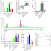
Figure 1:
Figure 1:. An. gambiae salivary apyrase activates tPA.
(A) Fluorogenic assay for single-chain tPA (sc-tPA) activation reveal that mosquito salivary gland extracts (SG) activate sc-tPA. Heating the extract at 65°C or 100°C prevents tPA activation. ****P <0.0001. (B) Fractions from An. gambiae salivary gland extracts obtained by size-exclusion chromatography were tested for tPA activation. Fraction Z8 showed higher sc-tPA activation compared to sc-tPA or SG alone. (C) An. gambiae salivary 5’ nucleotidase ecto (Ag5’NTE) identified as the saliva tPA activator. Ag5’NTE activates tPA at similar levels as An. gambiae saliva. **P <0.01. (D) Interaction of tPA with Ag5’NTE using ELISA overlays. rAg5’NTE and an unrelated protein (AGAP007393) were coated on the plate. Wells were incubated with or without sc-tPA. Anti-tPA antibodies were used to detect sc-tPA. Ag5’NTE interact with sc-tPA, whereas AGAP007393 does not interact (mustard yellow). A well was coated with sc-tPA as positive control. **P <0.01; ****P <0.0001. (E) Plasminogen activation in the presence of rAg5’ NTE and tPA measured by a colorimetric assay. Ag5’NTE activated tPA in-turn activates plasminogen to plasmin (dark pink) at higher levels than plasmin, sc-TPA or tc-tPA alone. **P <0.01; ****P <0.001; ns: not significant. (F) Ag5’NTE (AGAP011026) is an apyrase. Inorganic phosphate released was measured using Fiske Subbarow reagent. Ag5’NTE (AGAP011026), now referred as AgApyrase, released inorganic phosphate from ATP and ADP when compared to the positive control potato apyrase. ns: not significant. (G) rAgApyrase inhibits ADP-mediated platelet aggregation. Platelet-rich plasma was incubated with the platelet aggregation agonist ADP and platelet aggregation was measured by light transmittance over 6 min. rAgApyrase inhibited the platelet aggregation induced by ADP (orange) in contrast to the control buffer with ADP (blue) which aggregated all the available platelets. Data from three (A, D, E, F-ATP), two (C, F-ADP) or one (B) independent experiments. Groups were compared with an ordinary one-way ANOVA followed by Sidak’s multiple comparison test for pairwise comparison (A, C, D) or paired two-tailed t-test (E, F).
Figure 2:
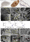
Figure 2:. Salivary AgApyrase is ingested during blood feeding and enhances fibrinolysis in the blood bolus.
(A) Immunohistochemistry performed on blood fed mosquito midgut shows ingestion of salivary AgApyrase in the mosquito midgut (dark brown patches in the middle panel) by staining with anti-apyrase antibodies. Blood fed mosquito midgut with no primary antibody did not show any signal (absence of brown patches in the first panel). Unfed mosquito midgut stained with anti-apyrase antibodies showed faint brown patches (black arrows in the third panel). MGE: midgut epithelium, MGL: midgut lumen. (B) Scanning electron microscopy (SEM) of blood boluses before and after supplementation with rAgApyrase. An. gambiae female mosquitoes were fed on mice before or after intravenous injection of rAgApyrase. Midguts were dissected at 30 min (or at 4 h shown in Fig. S8) post feeding. Note the well organize fibers formed in the blood bolus before the supplementation with rAgApyrase (white arrows) as compared to the thinner and sponge-like structure formed after rAgApyrase supplementation (yellow arrows). (C, D) Effect of rAgApyrase on D-dimer formation in blood boluses. Mosquitoes were fed on mice before or after intravenous injection of rAgApyrase, and midguts were dissected 30 min post feeding to measure D-dimer formation. Assays were done with pools of 5 (C) or 10 (D) midguts. Supplementation of the blood meal with rAgApyrase increases D-dimer formation. Data pooled from two independent experiments. Unpaired t-test, **P <0.002. (E) SEM of blood boluses from mosquitoes fed on rAgApyrase immunized mice. Control mosquitoes were fed on mice treated with adjuvant. Midguts were dissected 30 min post feeding. Note the increase in fiber organization and thickness (red arrows) observed in boluses from mosquitoes that fed on rAgApyrase immunized mice as compared to adjuvant treated mice.
Figure 3:
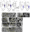
Figure 3:. Salivary AgApyrase inhibits platelet activation and aggregation in the blood bolus and decreases in vitro NETosis.
(A, B) Immunohistochemistry was performed on mosquito blood boluses before and after supplementation with rAgApyrase and stained with P-selectin (a marker for platelet activation). Quantification of the percentage area covered by P-selectin staining (A) and the mean P-selectin signal (B) per midgut (from IHC shown in Fig. S11). Data pooled from two independent experiments. Mann Whitney test, ***P=0.0001. (C, D) SEM showing platelet aggregation in blood boluses before and after supplementation with rAgApyrase (C) or in from mosquitoes fed on adjuvant or rAgApyrase immunized mice (D). White arrows indicate the aggregation of platelets containing filopodia. Yellow arrows show individual activated platelets that are not aggregated. Red asterisks show fragmentation of platelets into smaller vesicular bodies reminiscence of hypercoagulation. (E) rAgApyrase decreases in vitro NETosis in the presence of platelets. NETs were quantified by immunofluorescence microscopy with an anti-MPO antibody (Fig. S12A) and the percentage of NETs was calculated as an average of 5–10 fields (400X) normalized to total number of neutrophils. Results expressed as mean % ± SEM. Kruskal-Wallis Test, *P=0.01. (F, G) Immunohistochemistry as discussed in panel (A) was performed to quantify neutrophil elastase as a marker for NETosis. From IHC shown in Fig. S13. Data pooled from two independent experiments. Mann Whitney test, ****P=0.0001.
Figure 4:
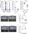
Figure 4:. Effect of AgApyrase on P. berghei transmission.
(A) AgApyrase facilitates P. berghei infection of mosquito midguts. Oocyst numbers from midguts of An. gambiae mosquitoes that fed on a P. berghei infected mouse before or after the intravenous injection of native or heat-denatured rAgApyrase. Data pooled from three individual experiments shown in Fig. S14, and groups were compared with two-tailed t-test followed by Mann-Whitney comparison test. Red lines indicate median. ****P <0.0001; *P<0.05. (B) AgApyrase immunization inhibits P. bergei midgut infection. Oocyst numbers were determined in the midguts of An. gambiae mosquitoes fed on P. berghei infected BALB/c mice previously immunized with rAgApyrase using Magic Mouse adjuvant or with adjuvant alone as control. Each dot represents median oocyst number from mosquitoes fed on one mouse. Groups were compared with two-tailed t-test followed by Mann-Whitney comparison test. Red lines indicate median. ****P <0.0001. The oocysts numbers from mosquitoes feeding on each individual mouse are shown on Fig. S15. (C) AgApyrase immunization reduces prevalence of Plasmodium midgut infection. The data obtained from the previous experiment was used to calculate the prevalence of infection. Each dot represents the prevalence of infected mosquitoes fed on one animal. Groups were compared with two-tailed t-test followed by Mann-Whitney comparison test. Red lines indicate median. ****P <0.0001. (D and E) AgApyrase immunization inhibits sporozoite transmission. BALB/c mice were immunized with rAgApyrase in Magic Mouse adjuvant or adjuvant alone (control). Mice were challenged with the bite of five An. stephensi mosquitoes infected with P. berghei sporozoites expressing the luciferase gene. Sporozoite infectivity was determined by measuring luciferase activity in the mouse liver 40 h post challenge. Luminescence signal in the mice livers is shown in panel D and the quantification in panel E. Data pooled from two independent experiments and groups were compared with two-tailed t-test followed by Mann-Whitney comparison test. Red lines indicate median. ***P <0.0002. (F and G) Similar experiment as in panels D and E, but mice were challenged with 2000 sporozoites injected intravenously. Data from a single experiment with five mice per group.

Similar articles

Cited by

References

    1. WHO, World malaria report 2022. 2022.
    1. Smith R.C., Vega-Rodriguez J., and Jacobs-Lorena M., The Plasmodium bottleneck: malaria parasite losses in the mosquito vector. Mem Inst Oswaldo Cruz, 2014. 109(5): p. 644–61. - PMC - PubMed
    1. Alves E.S.T.L., et al., The fibrinolytic system enables the onset of Plasmodium infection in the mosquito vector and the mammalian host. Sci Adv, 2021. 7(6). - PMC - PubMed
    1. Pascini T.V., et al., Transgenic Anopheles mosquitoes expressing human PAI-1 impair malaria transmission. Nat Commun, 2022. 13(1): p. 2949. - PMC - PubMed
    1. Pala Z.R., et al., Beyond cuts and scrapes: plasmin in malaria and other vector-borne diseases. Trends Parasitol, 2022. 38(2): p. 147–159. - PMC - PubMed

Publication types

LinkOut - more resources