[Preprint]. 2023 May 15:2023.05.15.540765.
doi: 10.1101/2023.05.15.540765.

A new antibiotic from an uncultured bacterium binds to an immutable target

Affiliations

A new antibiotic from an uncultured bacterium binds to an immutable target

Rhythm Shukla et al. bioRxiv. .

Update in

  • An antibiotic from an uncultured bacterium binds to an immutable target.
    Shukla R, Peoples AJ, Ludwig KC, Maity S, Derks MGN, De Benedetti S, Krueger AM, Vermeulen BJA, Harbig T, Lavore F, Kumar R, Honorato RV, Grein F, Nieselt K, Liu Y, Bonvin AMJJ, Baldus M, Kubitscheck U, Breukink E, Achorn C, Nitti A, Schwalen CJ, Spoering AL, Ling LL, Hughes D, Lelli M, Roos WH, Lewis K, Schneider T, Weingarth M. Shukla R, et al. Cell. 2023 Sep 14;186(19):4059-4073.e27. doi: 10.1016/j.cell.2023.07.038. Epub 2023 Aug 22. Cell. 2023. PMID: 37611581

Abstract

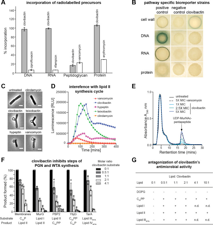
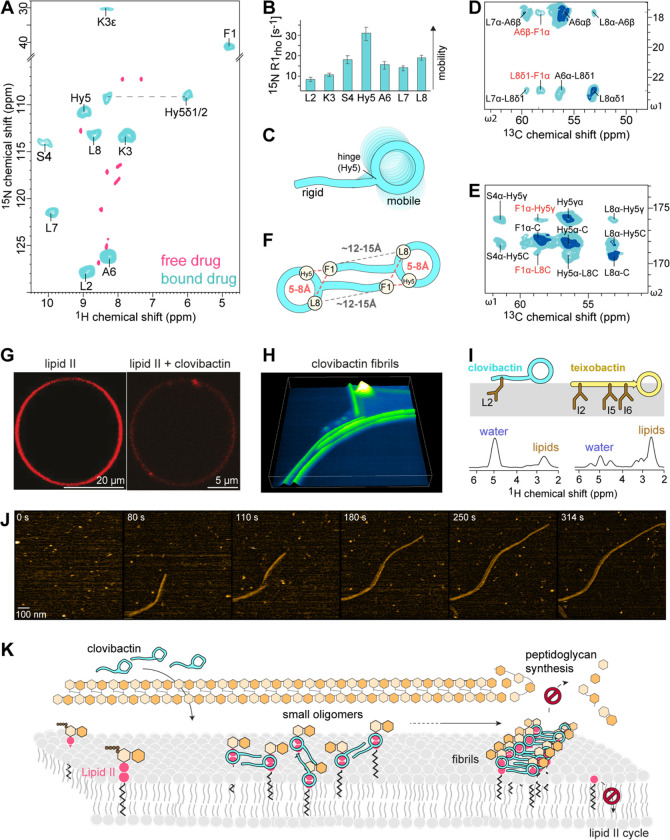
Antimicrobial resistance is a leading mortality factor worldwide. Here we report the discovery of clovibactin, a new antibiotic, isolated from uncultured soil bacteria. Clovibactin efficiently kills drug-resistant bacterial pathogens without detectable resistance. Using biochemical assays, solid-state NMR, and atomic force microscopy, we dissect its mode of action. Clovibactin blocks cell wall synthesis by targeting pyrophosphate of multiple essential peptidoglycan precursors (C 55 PP, Lipid II, Lipid WTA ). Clovibactin uses an unusual hydrophobic interface to tightly wrap around pyrophosphate, but bypasses the variable structural elements of precursors, accounting for the lack of resistance. Selective and efficient target binding is achieved by the irreversible sequestration of precursors into supramolecular fibrils that only form on bacterial membranes that contain lipid-anchored pyrophosphate groups. Uncultured bacteria offer a rich reservoir of antibiotics with new mechanisms of action that could replenish the antimicrobial discovery pipeline.


Conflict of interest statement

Competing Interests

The following authors, A. J. Peoples, C. Achorn, A. Nitti, A. L. Spoering, L. L. Ling, D. E. Hughes, and K. Lewis, declare competing financial interests as they are employees and consultants of NovoBiotic Pharmaceuticals. A patent US 11,203,616 B2 was issued 12/21/2021 and describes the use of clovibactin (Novo29) and as an antibiotic, as well as the pharmaceutical composition and antibiotic use of derivatives. The other authors have no competing interests.

Figures

Figure 1.
Figure 1.. Biosynthetic gene cluster and proposed biosynthesis of clovibactin.
The gene cluster associated with the biosynthesis of clovibactin was identified via whole genome sequencing and contained two nonribosomal peptide synthetases (NRPS) genes (cloA and cloB), a transporter gene (cloC) and a tailoring enzyme (cloD). Proposed biosynthetic pathway of clovibactin involves the assembly-line condensation of 8 canonical amino acids with 3 epimerizations carried out by dual-function condensation domains and a β-hydroxylation on Asn5 by the CloD, a TauD/TfdA dioxygenase. This hydroxylation provides the cyclization point for release from the NRPS and formation of the macrocyclic lactone.
Figure 2.
Figure 2.. Clovibactin kills S. aureus in vitro and in vivo.
(A) time-dependent killing of S. aureus (clovibactin (1xMIC), clovibactin (5xMIC), vancomycin (10xMIC) and chloramphenicol (5xMIC). (B,C) Clovibactin-induced lysis in S. aureus. Cells of S. aureus SA113 and a ΔaltA mutant were incubated with each compound at 2xMIC for 24 hours as indicated. Mean values from three independent experiments are shown. Error bars represent standard deviation. (D) Pharmacokinetic parameters of clovibactin in mice model determined using Watson LIMS software. (E) The bacterial load from the thigh infection model prior to dosing and 24 hours after treatment. The infection controls demonstrated a bioload of 6.07 log10 CFUs/ gram of thigh at the time of treatment (2 hours). Clovibactin was delivered as two IV doses (2 and 4 hours post infection) of 5, 10, 20 and 30 mg/kg. Vancomycin was delivered as a single IV dose at 50 mg/kg.
Figure 3.
Figure 3.. Clovibactin targets cell wall biosynthesis.
(A) Effect of clovibactin on macromolecular biosyntheses in S. aureus. Incorporation of 3H-thymidine (DNA), 3H-uridine (RNA), 3H-leucine (protein), and 3H-glucosamine (peptidoglycan) was determined in cells treated with clovibactin at 2xMIC (grey bars). Ciprofloxacin (8xMIC), rifampicin (4xMIC), vancomycin (2xMIC) and erythromycin (2xMIC) were used as positive controls (white bars). Data are averages of two independent experiments. (B) B. subtilis bioreporter strains with selected promotor-lacZ fusions were used to identify interference with major biosynthesis pathways. β-galactosidase (lacZ) is fused to promotors PypuA (cell wall), PyorB (DNA), PyvgS (RNA), and Pyhel (protein) and induction of a specific stress response is visualized by a blue halo at the edge of the inhibition zone. Antibiotics vancomycin, ciprofloxacin, rifampicin, and clindamycin were used as positive controls. (C) Clovibactin treatment results in cell-shape deformations and characteristic blebbing as observed by phase-contrast microscopy of B. subtilis. Cell wall active antibiotics teixobactin, hypeptin, vancomycin and protein synthesis inhibitor clindamycin were used as controls. Scale bar = 2 μm. (D) Clovibactin (1xMIC, blue) strongly induced Plial as observed by expression of the lux operon in B. subtilis PliaI-lux. Teixobactin, hypeptin, vancomycin and clindamycin were used as control antibiotics. (E) Intracellular accumulation of the soluble cell wall precursor UDP-MurNAc-pentapeptide after treatment of S. aureus with different concentrations of clovibactin. Untreated and VAN-treated (5xMIC) cells were used as controls. Experiments are representatives of 3 independent experiments. (F) Clovibactin inhibits membrane-associated steps of PGN and WTA synthesis in vitro. The antibiotic was added in molar ratios from 0.5 to 4 with respect to the amount of the lipid substrate C55P, C55PP, lipid II, or lipid IIIWTA used in the individual test systems. Reaction product synthesized in the absence of antibiotic was taken as 100%. Mean values from three independent experiments are shown. Error bars represent standard deviation. (G) Antagonization of the antimicrobial activity of clovibactin by cell wall precursors. S. aureus was incubated with clovibactin (8×MIC) in nutrient broth in microtiter plates, and growth was measured after a 24 h incubation at 37 °C. Putative HPLC-purified antagonists (undecaprenyl-pyrophosphate [C55PP], lipid I, lipid II, and lipid IIIWTA) and 1,2-dioleoyl-sn-glycero-3-phospho-glycerol (DOPG) were added in at molar ratios with respect to the antibiotic. Experiments were performed with biological replicates. +antagonization; - no antagonization.
Figure 4:
Figure 4:. High-resolution ssNMR structure and oligomerization of the clovibactin/lipid II complex in membranes.
(A) 2D NH ssNMR spectrum of lipid II bound clovibactin in membranes (cyan), superimposed on free clovibactin in aqueous solution (rose). (B) Site-resolved 15N R1rho dynamics of lipid II bound teixobactin and clovibactin in DOPC membranes. (C) Illustration of the NMR-derived dynamics. While clovibactin’s N-terminus is rigid, the depsi-cycle shows elevated dynamics. (D, E) Zooms into a 2D CC spectrum of the complex show head-to-tail contacts in clovibactin, suggesting a dimeric (supramolecular) arrangement of clovibactin in the complex. Data acquired at 1200 MHz magnetic field using 50 (cyan) and 250 ms (dark blue) CC mixing time. (F) Schematic illustration of the head-to-tail contacts between Phe1-Hyn5 and Phe1-Leu8 seen in (D, E) a dimeric model. (G) Confocal microscopy of DOPC GUVs doped with Atto-labelled lipid II and treated with clovibactin show domain/cluster formation. (H) 3D rendered high-speed AFM image show the formation of clovibactin – lipid II supramolecular fibrils after 10 minutes of interactions. (I) left: Mobility-edited (Doherty and Hong, 2009) ssNMR experiments show that clovibactin is much more water-accessible than teixobactin in the lipid II-bound state. right: ssNMR-derived topology and membrane insertion of clovibactin. (J) Snapshots of a timelapse HS-AFM video (Supplementary Video 1) following the assembly of clovibactin–lipid II fibrils. Images were obtained on a supported lipid bilayer containing 4% (mol) lipid II in the presence of 5 μM clovibactin, added at 0 s. Image acquisition rate of 0.5 frames per second. (K) Model of the mode of action of clovibactin. At the membrane surface, clovibactin binds lipid II and forms small oligomers that serve as nuclei for the formation of fibrils. Fibril formation enables a stable binding of lipid II and other cell wall precursors, blocking cell wall biosynthesis.
Figure 5:
Figure 5:. The interface and supramolecular structure.
(A) Chemical structure of lipid II. (B) 1D 31P ssNMR data in liposomes show marked changes of the lipid II PPi signal upon addition of clovibactin. 2D 1H31P ssNMR spectrum establishes direct interactions between the backbone of the depsi-cycle and PPi. (C) 2D CC ssNMR data of the 13C,15N-clovibactin – 13C,15N-lipid II complex in liposomes show interfacial contacts with the MurNAc sugar and the hydrophobic sidechains of the depsi-cycle. Interfacial contacts in blue. Data acquired at 950 MHz using 300 ms CC mixing time. (D) Sum of interfacial contacts with the sugars of lipid II. Shaded bars show ambiguous contacts of MurNAc with either K3 or L7. (E) Illustration of the interface: PPi and MurNAc are in direct proximity, GlcNAc is distal, the pentapeptide is flexible and not involved in the interface. (F) ssNMR-derived structural model of the clovibactin – lipid II complex. (G) Calculated interfaces of ssNMR structures superimpose very well and show that the hydrophobic depsi-cycle sidechains (Ala6, Leu7, Leu8) wrap like a glove around lipid II-PPi group, interacting with the hydrophobic side of MurNAc. (H) The cationic K3 and the polar S4 sidechains favorably interact with the lipid II PPi group. (I) Hydrophobic residues of clovibactin embrace the PPi-group like a glove, which appears entropically favourable by the release of boundary water.

References

    1. Baldus M., and Meier B.H. (1996). Total Correlation Spectroscopy in the Solid State. The Use of Scalar Couplings to Determine the Through-Bond Connectivity. Journal of Magnetic Resonance, Series A 121, 65–69.
    1. Breukink E., van Heusden H.E., Vollmerhaus P.J., Swiezewska E., Brunner L., Walker S., Heck A.J.R., and de Kruijff B. (2003). Lipid II is an intrinsic component of the pore induced by nisin in bacterial membranes. J Biol Chem 278, 19898–19903. - PubMed
    1. Brötz H., Bierbaum G., Leopold K., Reynolds P.E., and Sahl H.-G. (1998). The Lantibiotic Mersacidin Inhibits Peptidoglycan Synthesis by Targeting Lipid II. Antimicrobial Agents and Chemotherapy 42, 154–160. - PMC - PubMed
    1. Brown E.D., and Wright G.D. (2016). Antibacterial drug discovery in the resistance era. Nature 529, 336–343. - PubMed
    1. Buerger S., Spoering A., Gavrish E., Leslin C., Ling L., and Epstein S.S. (2012a). Microbial Scout Hypothesis and Microbial Discovery. Applied and Environmental Microbiology 78, 3229–3233. - PMC - PubMed

Publication types