[Preprint]. 2023 May 20:2023.05.19.541219.
doi: 10.1101/2023.05.19.541219.

CRISPR-based engineering of RNA viruses

Affiliations

CRISPR-based engineering of RNA viruses

Artem Nemudryi et al. bioRxiv. .

Update in

  • CRISPR-based engineering of RNA viruses.
    Nemudryi A, Nemudraia A, Nichols JE, Scherffius AM, Zahl T, Wiedenheft B. Nemudryi A, et al. Sci Adv. 2023 Sep 15;9(37):eadj8277. doi: 10.1126/sciadv.adj8277. Epub 2023 Sep 13. Sci Adv. 2023. PMID: 37703376 Free PMC article.

Abstract

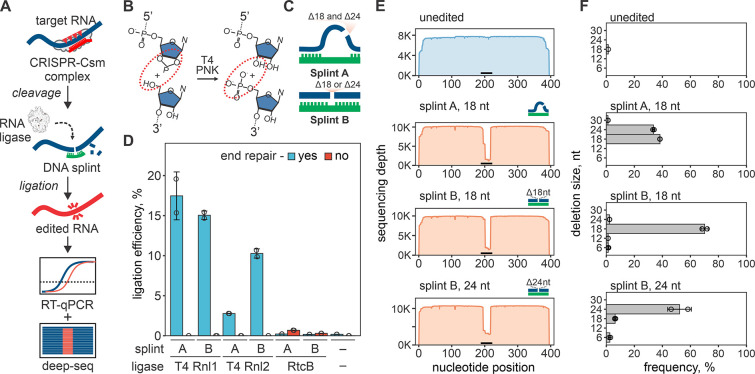
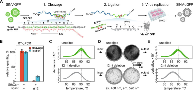
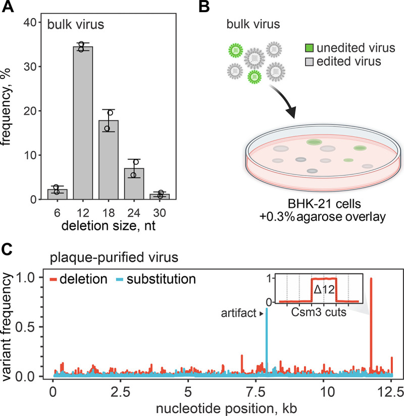
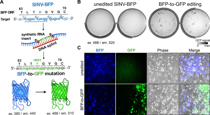
CRISPR RNA-guided endonucleases have enabled precise editing of DNA. However, options for editing RNA remain limited. Here, we combine sequence-specific RNA cleavage by CRISPR ribonucleases with programmable RNA repair to make precise deletions and insertions in RNA. This work establishes a new recombinant RNA technology with immediate applications for the facile engineering of RNA viruses.

One-sentence summary: Programmable CRISPR RNA-guided ribonucleases enable recombinant RNA technology.


Conflict of interest statement

Competing interests: B.W. is the founder of SurGene LLC and VIRIS Detection Systems Inc. B.W., A. Nemudryi, A. Nemudraia, and J.E.N. are inventors of the patent application US 17/811391 pertaining to use type III CRISPR-Cas system for sequence-specific editing of RNA viruses and gene therapy filed by Montana State University. The remaining authors declare no competing interests.

Figures

Fig. 1.
Fig. 1.. RNA editing with CRISPR-based cleavage and RNA ligation.
(A) Diagram of the RNA editing with type-III CRISPR nucleases and splinted ligation. Type-III CRISPR complexes cleave RNA in 6 nt intervals (red triangles), cutting out a portion of the target sequence. The resulting fragments are splint ligated to introduce edits. (B) SthCsm-mediated cleavage generates 2’3’-cyclic phosphate and 5’-hydroxyl ends (left, substrate for RtcB ligase) that can be converted to 3’-hydroxyl and 5’-phosphate (right, substrate for T4 RNA ligases) using T4 polynucleotide kinase (PNK). (C) Two splint designs. Hybridization of the DNA splint A to complementary RNA leaves single-stranded RNA flaps (curved blue lines) that imitate a break in the tRNA anti-codon loop, while splint B mimics nick in double-stranded RNA. (D) Comparison of RNA ligation efficiency with T4 RNA ligases (T4 Rnl) and RtcB ligase. Ligation efficiency was measured by performing RT-qPCR across the cut site and quantifying the signal relative to an uncut control (100%). (E) Deep sequencing of RNA ligated with splints depicted in (C). Horizontal black bar shows the target site of the SthCsm complex. (F) Quantification of editing outcomes in (E).
Fig. 2.
Fig. 2.. Programmed deletion of specific RNA sequence from the SINV-GFP genome.
(A) Diagram showing a pipeline for deleting 12 nucleotides from the GFP open reading frame (ORF) of the Sindbis-GFP (SINV-GFP) virus. See Fig. S2A for annotated genome map and Fig. S4A for additional experimental details. The deletion eliminates 4 codons encoding amino acids (L64, T65, Y66, G67) forming the chromophore of the fluorescent protein, which is expected to ablate fluorescence (“dead” GFP, dGFP). (B) RNA aliquots collected after the cleavage with the Csm complex (red) and ligation (blue) with T4 RNA ligase were reverse-transcribed and quantified with qPCR. Primers were designed to amplify cDNA across the target site. Relative quantities were calculated by normalizing to the uncut RNA control. (C) Melt curve analysis of the qPCR products generated with RNA after ligation in (B). Peaks indicate the melting temperature of the qPCR products. (D) BHK-21 cells were transfected with unedited or edited RNA of the SINV-GFP (two replicates each), seeded in 6-well plates, and imaged 24 hours later to capture the GFP signal (excitation 488 nm, emission 520 nm). (E) Melt curve analysis of the RT-qPCR products that were generated with RNA extracted from supernatants of BHK-21 cells 24 hours post-transfection. Primers were the same as in (B) and (C).
Fig. 3.
Fig. 3.. Plaque purification of edited viruses.
(A) Distribution of deletion sizes in sequencing reads that span the SthCsm target site in bulk edited virus. Data are shown as mean (n = 2) ± standard deviation. Dots show individual replicates. (B) Diagram of plaque purification approach used for isolating edited virus clones. (C) Frequency of nucleotide variants identified with amplicon sequencing of plaque purified virus. See Fig. S7 for details on sequencing strategy and analysis of other plaque-purified viruses. Black arrow indicates a sequencing artifact associated with amplicon-seq (see fig. S7). The inset is a close-up view of the targeted genomic region. Vertical dotted lines mark predicted cut sites of the SthCsm backbone subunit (Csm3) positioned in 6 nt intervals. The 12 nt deletion was intentionally selected using a DNA splint.
Fig. 4.
Fig. 4.. Programmed substitutions in viral genomes.
(A) Diagram showing editing strategy for substituting 12 nucleotides (underlined) in the blue fluorescent protein (BFP) open reading frame (ORF) of the recombinant Sindbis-BFP (SINV-BFP) virus. The C to U substitution at 11,762 recodes histidine residue for tyrosine (H66Y), which changes the fluorescence from blue to green (BFP-to-GFP). Additional silent substitutions (underlined, 11,761C>A and 11,764 C>U) were included to distinguish edited RNA from the SINV-GFP used for experiments shown in Fig. 2. (B) BHK-21 cells were transfected with the unedited or edited viral RNA (two replicates each), seeded on 6-well plates, and imaged after 24-hour incubation using 488 nm excitation laser and 520 nm emission filter. This laser and filter setting does not detect BFP (excitation 380 nm, emission 440 nm) in the unedited control, while replication of the edited BFP-to-GFP viruses is visible. (C) Transfected BHK-21 cells shown in (B) were imaged with an inverted fluorescent microscope at 20X magnification.

References

    1. Jackson D. A., Symons R. H., Berg P., Biochemical method for inserting new genetic information into DNA of Simian Virus 40: circular SV40 DNA molecules containing lambda phage genes and the galactose operon of Escherichia coli. Proceedings of the National Academy of Sciences 69, 2904–2909 (1972). - PMC - PubMed
    1. Berg P., Singer M. F., The recombinant DNA controversy: twenty years later. Proceedings of the National Academy of Sciences 92, 9011–9013 (1995). - PMC - PubMed
    1. Rouet P., Smih F., Jasin M., Introduction of double-strand breaks into the genome of mouse cells by expression of a rare-cutting endonuclease. Molecular and Cellular Biology 14, 8096–8106 (1994). - PMC - PubMed
    1. Stoddard B. L., Homing Endonucleases: From Microbial Genetic Invaders to Reagents for Targeted DNA Modification. Structure 19, 7–15 (2011). - PMC - PubMed
    1. Bibikova M. et al., Stimulation of homologous recombination through targeted cleavage by chimeric nucleases. Mol Cell Biol 21, 289–297 (2001). - PMC - PubMed

Publication types