[Preprint]. 2023 May 26:rs.3.rs-2879957.
doi: 10.21203/rs.3.rs-2879957/v1.

Lifetime of ground conformational state determines the activity of structured RNA

Affiliations

Lifetime of ground conformational state determines the activity of structured RNA

Rhese D Thompson et al. Res Sq. .

Update in

Abstract

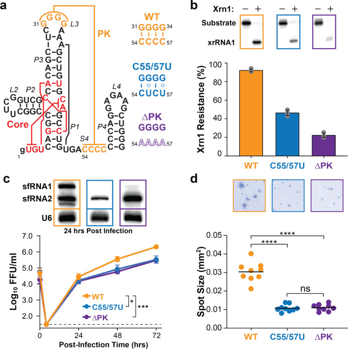
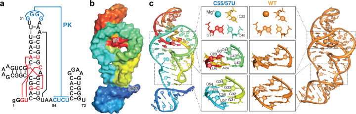
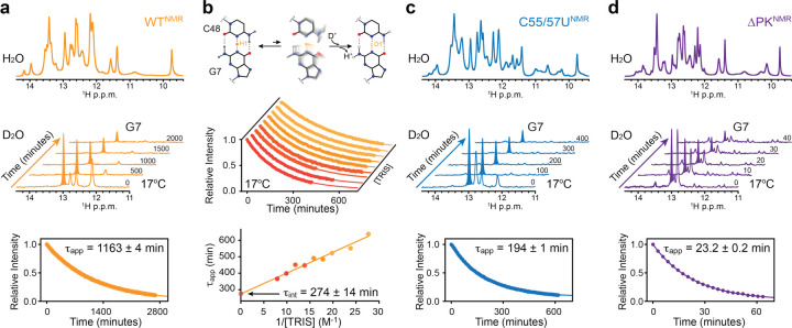
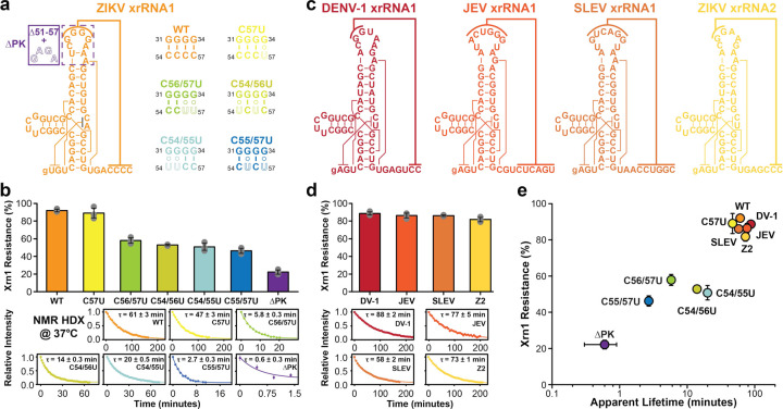
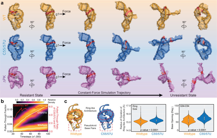
Biomolecules continually sample alternative conformations. Consequently, even the most energetically favored ground conformational state has a finite lifetime. Here, we show that, in addition to the 3D structure, the lifetime of a ground conformational state determines its biological activity. Using hydrogen-deuterium exchange nuclear magnetic resonance spectroscopy, we found that Zika virus exoribonuclease-resistant RNA (xrRNA) encodes a ground conformational state with a lifetime that is ~105-107 longer than that of canonical base pairs. Mutations that shorten the apparent lifetime of the ground state without affecting its 3D structure decreased exoribonuclease resistance in vitro and impaired virus replication in cells. Additionally, we observed this exceptionally long-lived ground state in xrRNAs from diverse infectious mosquito-borne flaviviruses. These results demonstrate the biological significance of the lifetime of a preorganized ground state and further suggest that elucidating the lifetimes of dominant 3D structures of biomolecules may be crucial for understanding their behaviors and functions.


Figures

Fig. 1 |
Fig. 1 |. Pseudoknot interaction modulates Xrn1 resistance of Zika virus xrRNA1.
a, Secondary structure of ZIKV xrRNA1 and pseudoknot sequences of wild-type (WT), weakening (C55/57U), and pseudoknot-less (DPK) mutants. b, In vitro Xrn1 digestion assays of WT, C55/57U, and DPK ZIKV xrRNA1s with 32P body labeling. Plotted are average percentages of Xrn1 resistance from three replicates with standard deviations as error bars. c, Quantification of viral replication of WT and mutant ZIKV in Vero cells. Viral titers were quantified using focus-forming assays and reported in the unit of focusforming units per milliliter (FFU/ml). Plotted are average values from 15–18 replicates from 5–6 independent experiments with standard error of the mean as error bars, where * and *** denote p-values <0.05 and <0.001, respectively, for statistical differences between WT and mutant ZIKV. Northern blots of sfRNAs from ZIKV-infected Vero cells are shown together with U6 RNA as an internal control. d, Quantification of cell-to-cell spread of WT and mutant ZIKV in Vero cells. Plotted are mean areas of infectious foci from 9 wells of 1 representative plate, where ns and **** denote p-values >0.05 and <0.0001, respectively, for statistical differences between WT and mutant ZIKV.
Fig. 2 |
Fig. 2 |. Structure of ZIKV xrRNA1s.
a, Secondary structure of the ZIKV xrRNA1 C55/57U construct in crystallography study. b, Electron density map, 2mFo-DFc, shown at 1σ on ZIKV xrRNA1 C55/57U construct, and surface representation of the crystal structure that highlights the ring architecture. c, Structural comparison between C55/57U (rainbow colored) and WT (orange, PDB: 5TPY) ZIKV xrRNA1s.
Fig. 3 |
Fig. 3 |. Conformational dynamics of ZIKV xrRNA1s by HDX NMR.
a, HDX NMR of WT xrRNA1 NMR construct at 17°C. Shown are 1H NMR spectra of the imino proton region in H2O and D2O. Time-dependent peak intensities of G7 imino proton upon dissolving in D2O are fit to a mono-exponential decay. Reported is the average apparent lifetime with s.d. estimated from fitting n = 241 data points. b, Tris-dependent HDX NMR of WT xrRNA1 NMR construct at 17°C. Shown is a schematic diagram that depicts the H-D exchange process of the G7 imino proton. Time-dependent peak intensities of G7 imino proton upon dissolving in D2O are plotted as a function of Tris concentration. Individual average apparent lifetimes with s.d. estimated from n = 49 to 100 data points are fit linearly to 1/[Tris] to extract the intrinsic lifetime of the G7 imino proton. c-d, HDX NMR of C55/57U (c) and DPK (d) xrRNA1 NMR constructs at 17°C. Shown are 1H NMR spectra of the imino proton region in H2O and D2O. Time-dependent peak intensities of G7 imino proton upon dissolving in D2O are fit to a mono-exponential decay. Reported are average apparent lifetimes with s.d. estimated from fitting n = 215 and 23 data points for C55/57U and DPK, respectively.
Fig. 4 |
Fig. 4 |. Conformational dynamics and Xrn1 resistance of MBFV xrRNAs.
a, Secondary structures of WT and mutant ZIKV xrRNA1s. b, Dynamic and functional analyses of WT and mutant ZIKV xrRNA1s at 37°C. Shown are the secondary structure of ZIKV xrRNA1 and pseudoknot sequences of WT and mutants. Plotted are average percentages of Xrn1 resistance from three replicates of in vitro Xrn1 digestion assay with standard deviations as error bars. Time-dependent peak intensities of G7 imino proton upon dissolving in D2O are plotted for each construct and fit to mono-exponential decays. Reported are average apparent lifetimes with s.d. estimated from fitting two independent measurements with a total of n = 59, 40, 27, 92, 94, 27, 4 data points for WT, C57U, C56/57U, C54/56U, C54/55U, C55/57U, and DPK, respectively. c, Secondary structures of other mosquito-borne flavivirus xrRNAs. d, Dynamic and functional analyses of other mosquito-borne flavivirus xrRNAs at 37°C. Plotted are average Xrn1 resistances of dengue virus 1 xrRNA1 (DENV-1), Japanese encephalitis virus xrRNA1 (JEV), Saint Louis encephalitis virus xrRNA1 (SLEV), and ZIKV xrRNA2 (Z2) from three replicates of in vitro Xrn1 digestion assay with standard deviations as error bars. Time-dependent peak intensities of G7 imino proton upon dissolving in D2O are plotted for each construct and fit to mono-exponential decays. Reported are average apparent lifetimes with s.d. estimated from fitting two independent measurements with a total of n = 82, 66, 65, 74 data points for DENV-1, JEV, SLEV, and Z2, respectively. e, Diagram of Xrn1 resistances and the apparent lifetimes of G7 imino proton of xrRNAs.
Fig. 5 |
Fig. 5 |. Xrn1 resistance of ZIKV xrRNA1s via constant-force simulations.
a, Representative trajectories of WT, C55/57U, and DPK ZIKV xrRNA1s from constantforce molecular dynamics (CFMD) simulations at 400 pN. Shown are states of xrRNA1s captured at the same timesteps along trajectories. The conserved G7–C48–C22 triple is highlighted in red. b, Accumulated pulled-through fractions of WT and C55/57U xrRNA1s as a function of timesteps in CFMD simulations. The schematic diagram depicts a pulled-through event as a disrupted G7–C48–C22 triple. c, Distribution of the sizes of the ring architecture of WT and C55/57U xrRNA1s in CFMD simulations. Plotted are receiver operating characteristic (ROC) analyses of the ring size and pulledthrough event from WT (orange), C55/57U (blue), and combined (black) trajectories with corresponding area under the curve (AUC) values. Distribution of the opening degrees of the pseudoknot base pair G34–C54 of WT and C55/57U xrRNA1s in CFMD simulations. Plotted are ROC analyses of the base opening and the ring architecture opening from WT (orange), C55/57U (blue), and combined (black) trajectories with corresponding AUC values.
Fig. 6 |
Fig. 6 |. Model for xrRNA-dependent Xrn1 resistance.
xrRNA acts as a molecular wall to inhibit Xrn1 digestion towards generating subgenomic flavivirus RNAs to promote viral replication. The strength of exonuclease resistance is influenced by the lifetime of the ground conformational state (GS) of the closed base pair anchored on the ring architecture, which is modulated through the long-range pseudoknot interaction.

References

    1. Henzler-Wildman K. & Kern D. Dynamic personalities of proteins. Nature 450, 964–972 (2007). - PubMed
    1. Ganser L. R., Kelly M. L., Herschlag D. & Al-Hashimi H. M. The roles of structural dynamics in the cellular functions of RNAs. Nat. Rev. Mol. Cell Biol. 20, 474–489 (2019). - PMC - PubMed
    1. Alderson T. R. & Kay L. E. NMR spectroscopy captures the essential role of dynamics in regulating biomolecular function. Cell 184, 577–595 (2021). - PubMed
    1. Boehr D. D., McElheny D., Dyson H. J. & Wright P. E. The dynamic energy landscape of dihydrofolate reductase catalysis. Science 313, 1638–1642 (2006). - PubMed
    1. Fraser J. S. et al. Hidden alternative structures of proline isomerase essential for catalysis. Nature 462, 669–673 (2009). - PMC - PubMed

Publication types

LinkOut - more resources