[Preprint]. 2023 May 16:rs.3.rs-2913647.
doi: 10.21203/rs.3.rs-2913647/v1.

Parallelized immunomagnetic nanopore sorting: modeling, scaling, and optimization of surface marker specific isolation of extracellular vesicles from complex media

Affiliations

Parallelized immunomagnetic nanopore sorting: modeling, scaling, and optimization of surface marker specific isolation of extracellular vesicles from complex media

Andrew A Lin et al. Res Sq. .

Update in

Abstract

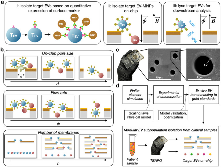
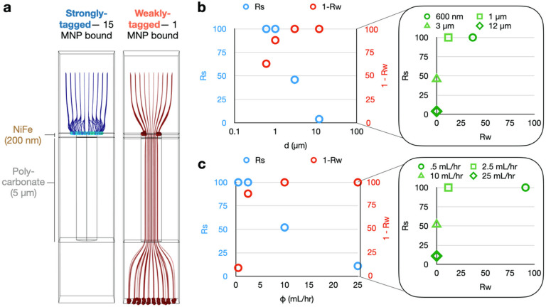
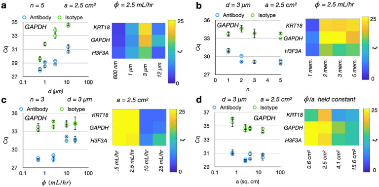
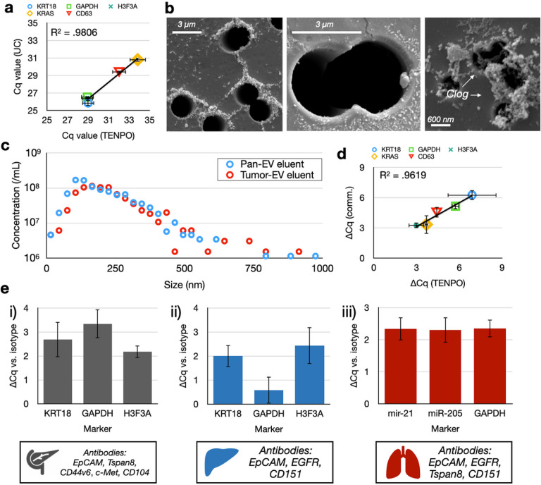
The isolation of specific subpopulations of extracellular vesicles (EVs) based on their expression of surface markers poses a significant challenge due to their nanoscale size (< 800 nm), their heterogeneous surface marker expression, and the vast number of background EVs present in clinical specimens (10 10 -10 12 EVs/mL in blood). Highly parallelized nanomagnetic sorting using track etched magnetic nanopore (TENPO) chips has achieved precise immunospecific sorting with high throughput and resilience to clogging. However, there has not yet been a systematic study of the design parameters that control the trade-offs in throughput, target EV recovery, and specificity in this approach. We combine finite-element simulation and experimental characterization of TENPO chips to elucidate design rules to isolate EV subpopulations from blood. We demonstrate the utility of this approach by increasing specificity > 10x relative to prior published designs without sacrificing recovery of the target EVs by selecting pore diameter, number of membranes placed in series, and flow rate. We compare TENPO-isolated EVs to those of gold-standard methods of EV isolation and demonstrate its utility for wide application and modularity by targeting subpopulations of EVs from multiple models of disease including lung cancer, pancreatic cancer, and liver cancer.


Conflict of interest statement

Conflicts of Interest

For our conflicts of interest to disclose, Dr. David Issadore is a founder of Chip Diagnostics and holds equity in the company. The other authors listed do not have competing interests.

Figures

Figure 1
Figure 1
Characterization of TENPO isolation of EV subpopulations. (A) Schematic of track-etched magnetic nanopore EV isolation. EVs are first labeled with biotinylated capture antibodies followed by anti-biotin magnetic nanoparticles (50 nm). EV-MNP complexes are magnetically captured as they flow vertically through parallelized magnetic nanopores. (B) Illustrations of tradeoffs in TENPO isolation. Adjusting the design parameters - pore diameter d, flow rate, and number of membranes n - results in trade-offs that can be used to tailor TENPO to isolate particular EV subpopulations from clinical specimens.(C) Photograph of an assembled TENPO chip (left) and SEM micrographs of the TENPO magnetic nanopores (center and right) with an EV immobilized on-chip (right). (D) A schematic of the workflow of this study.
Figure 2
Figure 2
Finite-element simulations to characterize TENPO EV sorting. (A) Particle tracking simulations for strongly-tagged versus weakly-tagged EVs through a single magnetic nanopore at an example pore diameter d = 1 μm and an example volumetric flow rate = 2.5 mL/hr. (B) The capture rate of strongly labeled EVs (Rs) and weakly labeled EVs (Rw) versus pore diameter d for a volumetric flow rate = 2.5 mL/hr. (C) The capture rate of strongly labeled EVs and weakly labeled EVs versus volumetric flow rate, for a pore diameter d = 1 μm.
Figure 3
Figure 3
Experimental characterization of TENPO isolation in an in vitro model system of pancreatic cancer. Device parameters which were held constant in the course of the parameter scan are labeled atop each graph set. Each dot represents one device replicate, and error bars are from n = 2 PCR replicates; each condition was ran with two antibody device replicates and two isotype device replicates. Fold-change enrichment ζ was calculated for each condition as 2^(ΔCq) for the ΔCq between antibody versus isotype devices. (A) Isolated EV RNA as a function of pore diameters d for antibody-labeled versus isotype-labeled EVs. (B) Isolated EV RNA as a function of membrane number n for antibody-labeled versus isotype-labeled EVs. (C) Isolated EV RNA as a function of flow rate for antibody-labeled versus isotype-labeled EVs. (D) Isolated EV RNA as a function of cross-sectional area a for antibody-labeled versus isotype-labeled EVs.
Figure 4
Figure 4
In vitro benchmarking of TENPO to gold standard technologies and in different biological systems. (A) Correlation of nucleic acid cargoes between TENPOvs. UC. Each point corresponds to a nucleic acid marker measured in the CD9/CD63/CD81+ EVs isolated using TENPO compared to the same nucleic acid markers measured in EVs isolated using UC. Error bars from n = 2 device/prep replicates. (B) SEM micrographs of EVs captured on TENPO. The left and middle micrographs show a TENPO with d = 3 μm magnetic nanopores. The right micrograph shows a clogged TENPO with d = 600 nm magnetic nanopores. (C) Size distributions of EVs captured by TENPO and eluted for measurement by NTA. (D) Comparison of ΔCq between cancer cell culture media spiked into plasma versus control cell culture media spiked into plasma for pan-EV TENPO vs. a commercial pan-EV kit. Error bars from n = 2 device/prep replicates (two case, two control) using propagation of error. (E) Comparison of ΔCq between antibody-labeled versus isotype-control-labeled EVs in three different model systems of cancer. Error bars from n = 2 device/prep replicates (two antibody devices, two isotype devices) using propagation of error.

References

    1. Yu W. et al. Exosome-based liquid biopsies in cancer: opportunities and challenges. Annals Oncology. 32(4), 466–477 (2021). - PMC - PubMed
    1. Kumar S. R. et al. RNA cargos in extracellular vesicles derived from blood serum in pancreas associated conditions. Sci. Rep. 10, 2800 (2020). - PMC - PubMed
    1. Musante L. et al. Rigorous characterization of urinary extracellular vesicles (uEVs) in the low centrifugation pellet - a neglected source for uEVs. Sci. Rep. 10, 3701 (2020). - PMC - PubMed
    1. Lin A. A., Nimgaonkar V., Issadore D., & Carpenter E. L. Extracellular Vesicle–Based Multianalyte Liquid Biopsy as a Diagnostic for Cancer. Ann. Rev. of Biomed. Data Sci, 5. (2022). - PubMed
    1. Ko J., et al. Combining machine learning and nanofluidic technology to diagnose pancreatic cancer using exosomes. ACS Nano, 11(11), 11182–11193. (2017). - PubMed

Publication types

LinkOut - more resources