[Preprint]. 2023 May 15:rs.3.rs-2917340.
doi: 10.21203/rs.3.rs-2917340/v1.

Mesenchymal-Derived Extracellular Vesicles Enhance Microglia-mediated Synapse Remodeling after Cortical Injury in Rhesus Monkeys

Affiliations

Mesenchymal-Derived Extracellular Vesicles Enhance Microglia-mediated Synapse Remodeling after Cortical Injury in Rhesus Monkeys

Yuxin Zhou et al. Res Sq. .

Update in

Abstract

Understanding the microglial neuro-immune interactions in the primate brain is vital to developing therapeutics for cortical injury, such as stroke. Our previous work showed that mesenchymal-derived extracellular vesicles (MSC-EVs) enhanced motor recovery in aged rhesus monkeys post-injury of primary motor cortex (M1), by promoting homeostatic ramified microglia, reducing injury-related neuronal hyperexcitability, and enhancing synaptic plasticity in perilesional cortices. The current study addresses how these injury- and recovery-associated changes relate to structural and molecular interactions between microglia and neuronal synapses. Using multi-labeling immunohistochemistry, high resolution microscopy, and gene expression analysis, we quantified co-expression of synaptic markers (VGLUTs, GLURs, VGAT, GABARs), microglia markers (Iba-1, P2RY12), and C1q, a complement pathway protein for microglia-mediated synapse phagocytosis, in perilesional M1 and premotor cortices (PMC) of monkeys with intravenous infusions of either vehicle (veh) or EVs post-injury. We compared this lesion cohort to aged-matched non-lesion controls. Our findings revealed a lesion-related loss of excitatory synapses in perilesional areas, which was ameliorated by EV treatment. Further, we found region-dependent effects of EV on microglia and C1q expression. In perilesional M1, EV treatment and enhanced functional recovery were associated with increased expression of C1q + hypertrophic microglia, which are thought to have a role in debris-clearance and anti-inflammatory functions. In PMC, EV treatment was associated with decreased C1q + synaptic tagging and microglial-spine contacts. Our results provided evidence that EV treatment facilitated synaptic plasticity by enhancing clearance of acute damage in perilesional M1, and thereby preventing chronic inflammation and excessive synaptic loss in PMC. These mechanisms may act to preserve synaptic cortical motor networks and a balanced normative M1/PMC synaptic connectivity to support functional recovery after injury.


Conflict of interest statement

Competing interests: The authors declare that they have no competing interests.

Figures

Figure 1
Figure 1. Experimental design and representative Images of immunolabeled markers, lesion, and sampling location.
a Experimental workflow: monkeys were trained and tested on a fine motor task – the Hand Dexterity Task, before and after the surgical lesion of M1 as described in Moore et al., 2019. Then, the monkeys were randomly assigned with either vehicle or EV treatment, which were infused IV at 24 hours and 14 days post-injury. The brains were harvested 14 to 16 weeks after the surgery. (a1) During Krebs buffer perfusion, 1–2cm fresh tissue block was harvested from the ventral perilesional motor/premotor cortex: the caudal 1/4 was processed for qPCR and the rostral part was cut into 300 μm acute slices for whole cell patch clamp recording and intracellular filling of layer 3 pyramidal cells. (a2) The remainder of the brain containing the lesion and rostral and dorsal perilesional cortex was fixed with paraformaldehyde then cut into serial coronal sections and processed with immunohistochemical labeling. The synaptic markers that were immuno-labeled included: VGLUT1 &2, GLUR2/3, VGAT, GABAa α1, and GABAb R2. Microglia markers were combining labeled Iba-1 and P2RY12. C1q was immuno-labeled to indicate complement activation. Pyramidal neurons were intracellularly filled with internal solution with 1% biocytin. b Sample image of surgical lesion and sampling location: perilesional primary motor (M1) and premotor cortices (PMC)
Figure 2
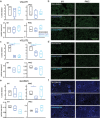
Figure 2. The expression of excitatory synaptic markers in perilesional M1 and PMC.
a The density (% area labeled) and average particle size (area in μm2) of VGLUT1+ puncta in perilesional M1 and in PMC. Non-lesion control: n=3. Veh group: n=4. EV group: n=5. b Representative maximum-projection confocal images of VGLUT1 immuno-labeling in M1 and PMC. Scale bar: 20μm. c The density and average size of VGLUT2+ puncta in perilesional M1 and in PMC. The density of VGLUT2+ puncta was significantly lower in groups with lesions compared with non-lesion controls (con) in both perilesional M1 (Fisher’s LSD post hoc, con. vs veh: p=0.004; con vs EV: p=0.001) and in PMC (Fisher’s LSD post hoc, con. vs veh: p=0.04; con vs EV: p=0.02). Non-lesion control: n=3. Veh group: n=4. EV group: n=5. d Representative maximum-projection confocal images of VGLUT2 immuno-labeling in M1 and PMC. Scale bar: 20μm. e The density (% area label) and average size (μm2) of GluR2/3+ puncta in perilesional M1 and in PMC. The density of GLUR2/3+ puncta in PMC was significantly lower in the vehicle group (Fisher’s LSD post hoc, con. vs veh, p=0.036) but not in the EV group (Fisher’s LSD post hoc, con. vs EV, p=0.49) as compared with non-lesion controls. Non-lesion control: n=3. Veh group: n=4. EV group: n=4. f Representative maximum-projection confocal images of GLUR2/3 immuno-labeling in M1 and PMC. Scale bar: 20μm
Figure 3
Figure 3. The expression of inhibitory synaptic markers in perilesional M1 and PMC.
a The density and average size of VGAT in perilesional M1 and in PMC. No significant result was found. Non-lesion control: n=3. Veh group: n=5. EV group: n=5. b Representative maximum-projection confocal images of VGAT immuno-label in M1 and PMC. Scale bar: 20μm. c. The density and average size of GABAa α1 in perilesional M1 and in PMC. The density of GABAa α1 subunit in perilesional M1 was significantly lower in both veh (Fisher’s LSD post hoc, con. vs. Veh, p=0.038) and EV group (Fisher’s LSD post hoc, con. vs. EV, p=0.009) as compared with non-lesion controls. The density of GABAa α1 in PMC was lower in veh (Fisher’s LSD post hoc, con. vs. veh, p=0.05) and EV group (Fisher’s LSD post hoc, con. vs. EV, p=0.02). The size of GABAa α1 in M1 was significantly smaller in the EV group (Fisher’s LSD post hoc, con. vs. EV, p<0.001). Non-lesion control: n=3. Veh group: n=4. EV group: n=5. d Representative maximum-projection confocal images of GABAa α1 receptor subunits immuno-label in M1 and PMC. Scale bar: 20μm. e. The density and particle average size of GABAb R2 subunit in perilesional M1 and in PMC. Non-lesion control: n=3. Veh group: n=4. EV group: n=5. f Representative maximum-projection confocal images of and GABAb R2 subunits immuno-label in M1 and PMC. Scale bar: 20μm
Figure 4
Figure 4. Glutamate and GABA receptors subunit mRNA expression in perilesional cortex.
a Fold changes of glutamate receptor subunit gene expression. The gene expression of GRIA2 (GluR2) was significantly higher in the veh group, as compared with non-lesion controls (t-test, p < 0.001) and the EV group (t-test, p=0.01). b Fold changes of GABA receptor subunit gene expression. The gene expression of GABRD (GABAa ∂) was significantly higher in the EV group as compared with non-lesion controls (t-test, p=0.004). Gene names: GRIA1 (AMPA GluR1), GRIA2 (AMPA GLuR2), GRIN1 (NMDA NR1), GRIN2B (NMDA NR2B), GABRA1 (GABAa α1), GABRA2 (GABAa α2), GABRA5 (GABAa α5), GABRD (GABAa ∂), GABBR2 (GABAb R2). c MDS plot showing clustering of cases based on mRNA expression profiles of Glu and GABA receptor subunits. The proximity of points indicates the relative similarity-based pair-wise correlation of these multiple mRNA expression variables
Figure 5
Figure 5. Microglia apposition on synaptic structures.
a Representative images of microglia interactions with dendritic spines/shaft at different z-levels of stack. Neuronal dendrites and spines were filled with biocytin and stained with Streptavidin-Alexa 488 (green) and microglia were visualized with Iba-1 & P2RY12 + (red) immuno-labeling. White pixels indicate an overlap between two channels. Scale bar: 20μm and 5μm. b Schematic diagram of criteria for determining microglia appositions on dendritic shafts or spines, classified as either contact (touching) or neighboring (within 1 microns). c. The density of microglia appositions (contacting & neighboring) on dendritic shaft and spine. The vehicle group had higher densities of microglial-apical shaft (Fisher’s LSD post hoc, veh vs. EV, p=0.009) and microglial-apical spine (p=0.038) appositions compared to EV, and a trend for greater total appositions in the vehicle than in the control was found (con. vs. veh., p=0.058). d Total microglial appositions (contacts & neighboring on spines and shafts) on different segments of apical/basal dendrites in vPMC. Mid-apical dendrites had higher density of microglial contacts only in veh compared to control (Fisher’s LSD post hoc, con. vs veh, p=0.03; con. vs EV, p=0.84). Non-lesion control: n=10 from 3 monkeys. Veh group: n=10 from 3 monkeys. EV group: n=7 from 2 monkeys. e The fraction of VGLUT2 colocalized with microglia was higher in both groups with lesions in M1 (Fisher’s LSD post hoc, con. vs. veh, p=0.009; con. vs. EV, p<0.001). Non-lesion control: n=3. Veh group: n=4. EV group: n=5. f Representative images of dual channel labeling (left panel) of microglia (red) and VGLUT2 (green), with right panel showing higher resolution of colocalized VGLUT2-microglia label masked in white
Figure 6
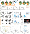
Figure 6. C1q co-expression on VGLUT2+ axon terminals and Iba1 microglia.
a The density (% area label) of C1q+ puncta in perilesional M1 and PMC (Fisher’s LSD post hoc, M1: con. vs EV p=0.028; Non-lesion control: n=5. Veh group: n=5. EV group: n=4). b Fold changes of C1QA and C3 gene expression in perilesional M1 (t-test, veh. vs. EV, p=0.027). c The density (% area label) of VGLUT2+ puncta colocalized with C1q+ puncta (Fisher’s LSD post hoc, con. vs. veh, p=0.02; con. vs. EV, p=0.02; Non-lesion control: n=3. Veh group: n=4. EV group: n=5). d Representative maximum-projection confocal images of C1q+ puncta immuno-labeling in M1 and PMC. Scale bar: 20μm. e Representative maximum-projection confocal images of 4 optical slices showing dual channel labeling (left panel) of C1q (yellow) and VGLUT2 (green), with colocalized VGLUT2–C1q points masked in white (right panel). Scale bar: 20μm. f The distance between the C1q-VGLUT2 colocalized points and microglia (Iba1)-VGLUT2 colocalized points. Inset shows schematic diagram of how C1q-VGLUT2-Iba1 distance was determined. Non-lesion control: n=3. Veh group: n=4. EV group: n=5. g Linear regression showing increasing C1q expression correlated with decreasing expression of VGLUT2+ (R2 = 0.378, p=0.034) in M1. h Linear regression greater C1q expression correlated with greater VGLUT2-Microglia colocalization (R2 = 0.372, p=0.035). i Representative maximum-projection confocal images of 9 optical slices showing single/dual/triple channel labeling (left to right) of colocalized VGLUT2-Iba1–C1q
Figure 7
Figure 7. C1q expression in different microglia phenotypes.
a The density (% area label) of Iba1+ colocalized with C1q+ puncta (Fisher’s LSD post hoc, M1: con. vs. veh, p=0.046; con. vs. EV, p=0.003; veh vs. EV, p=0.009); PMC: con. vs. EV p=0.004. Non-lesion control: n=3. Veh group: n=4. EV group: n=5). b Higher fraction of VGLUT2 colocalized with microglia (Iba1) in EV than in the veh. (Fisher’s LSD post hoc, veh. vs. EV: p=0.05. Non-lesion control: n=3. Veh group: n=4. EV group: n=5). cRepresentative images of dual labeling of microglia (red) and C1q (yellow), with examples of different microglia morphologies dual labeled with C1q. dCell densities of total C1q+ vs C1q- microglia. In M1, C1q- microglia: greater density in lesion than control (t-test, con. vs. veh: p<0.001; con. vs. EV: p=0.004); EV but not veh had greater C1q+ microglia density than control (t-test, con. vs. veh: p=0.07; con. vs. EV: p=0.03). In PMC, greater density of C1q+ microglia in veh than control group (t-test, con. vs. veh: p=0.02). e In M1, hypertrophic microglia: greater density in lesion compared to control group (t-test, con. vs. veh: p=0.01; con. vs. EV: p=0.008). f In M1, the EV group showed higher density of C1q- ramified microglia as compared to the control group (t-test, con. vs. EV: p=0.01). Both groups with lesion had higher density of hypertrophic C1q+ microglia as compared to the controls (t-test, con. vs. veh: p=0.047; con. vs. EV: p<0.001). However, only veh had higher density of hypertrophic C1q- microglia as compared to the controls (t-test, con. vs. veh: p=0.005). g In PMC, no between-group difference was found in the density of ramified or hypertrophic microglia. (d-g non-lesion control: n=3. Veh group: n=4. EV group: n=5)
Figure 8
Figure 8. The expression of different microglia phenotypes in M1 and PMC.
a Pie charts of %microglia by morphology and C1q expression in M1. Right inset shows the ratio of Hyper+ to the total Hyper microglia in each group (t-test, con. vs. veh: p=0.001; con. vs. EV: p=0.03; veh. vs. EV: p=0.006). b In PMC (Rami-veh < con.: p=0.05; EV vs con.: p=0.49). c Relative proportion of Rami+, Hyper+, Rami-, and Total C1q+ normalized to the total number of microglia counted for each case. Significant group*area interaction for %Rami+ (p=0.008), %Hyper- (p=0.02), and %Total C1q+ (p=0.04). A main effect of group for %Hyper+ (p=0.01). Significant between-group differences per area: M1 (%Rami+ veh < con, p=0.006; EV vs con, p=0.004; %Hyper+, EV > con, p=0.01; %Hyper- veh. > con. p=0.007). PMC (%C1q+ veh > con, p=0.02). Significant between-area differences per group: Control (%Rami+ M1 > PMC, p=0.009; and %C1q+ M1 > PMC, p=0.02). Vehicle (%Rami+: PMC > M1, p=0.05; but %Hyper-: M1 > PMC, p=0.01). Non-lesion control: n=3. Veh group: n=4. EV group: n=5. dRepresentative microglia reconstructions. e-g Morphological parameters (non-lesion control: n=36 cells from 1 monkey. veh group: n=58 cells from 1 monkey. EV group: n=71 cells from 1 monkey): e Number of primary process (three-way ANOVA, main effect ‘morphology’: p=0.02; ‘group’: p=0.01). Hyper+ (Fisher’s LSD post-hoc EV > con, p=0.03; EV > veh, p = 0.05); Hyper- (EV > veh, p = 0.02); Rami+ (con < veh, p=0.03; con < EV, p=0.05); EV group (Hyper+ > Rami+: p=0.004; Hyper- > Rami-: p<0.001). f Microglia soma surface area (three-way ANOVA, main effect ‘group’: p=0.006; ‘C1q+/−’: p=0.004). Hyper+: EV > con., p=0.01; Rami+: EV > con, p=0.04. g Soma aspect ratio (three-way ANOVA, main effect ‘C1q+/−’: p=0.03). h 3D scatter plot of morphological parameters. Annotations based on morphology, C1q expression, and experimental group
Figure 9
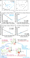
Figure 9. Relationship of synaptic and microglia properties to behavior outcome features.
a NMDS plot showing clustering of cases, annotated by experimental group (left) and cortical area (right), based on 21 synaptic and microglia outcome measures (%area VGLUT1, VGLUT2, VGAT, GLUR2/3, GABAA alpha1, GABAB R2; % of VGLUT1, VGLUT2 or VGAT with Iba1; % of IBA1 with VGLUT1, VGLUT2 or VGAT; % area C1q; % of VGLUT2 with C1q; % C1q with VGLUT2; cell densities of Ramified, Hypertrophic, Amoeboid C1q+ and C1q- microglia). The proximity of points indicates the relative similarity-based pair-wise correlation of these multiple variables. b-e Significant linear correlations between synaptic-microglial measures and behavioral outcome measures: b Increased density of C1q and Iba1 colocalization in M1 correlated with faster recovery time (less days to return to pre-operative latency to retrieve food reward; R2 = 0.752, p=0.002). c Increased fraction of VGLUT2 colocalized with C1q in PMC correlated with slower recovery time (more days return to preoperative grasp pattern; R2 = 0.589, p=0.016). d Greater expression of C1q+ hypertrophic microglia in M1 was correlated with faster recovery time (R2 = 0.533, p=0.026). d Greater expression of C1q+ ramified microglia in PMC was associated with slower recovery time (R2 = 0.490, p=0.036). f A schematic showing summary of findings and proposed model of the lesion and EV treatment effects on microglial-synapse modulation and C1q signaling pathways. Cortical lesion in M1 induces acute damaged in neuronal structures that triggers an acute increase in C1q+ signaling cascade to initiate phagocytotic clearance. The veh group had accumulation of further damage and downstream C1q pathway related proteins that sustains a chronic pro-inflammatory response (C1q- hypertrophic microglia). The EV treatment upregulated C1q+ mediated clearance of debris and facilitated an early shift to the anti-inflammatory C1q+ hypertrophic microglia phenotype that persisted in the chronic stages, thereby supporting functional recovery

Similar articles

References

    1. Winship IR, Murphy TH. Remapping the Somatosensory Cortex after Stroke: Insight from Imaging the Synapse to Network. Neuroscientist. SAGE Publications Inc STM; 2009;15:507–24. - PubMed
    1. Edwards I, Singh I, Rose’meyer R. The Role of Cortisol in the Development of Post-Stroke Dementia: A Narrative Review. Heart Mind. 2022;6:151.
    1. Carmichael ST. Brain Excitability in Stroke: The Yin and Yang of Stroke Progression. Archives of Neurology. 2012;69:161–7. - PMC - PubMed
    1. Patel AR, Ritzel R, McCullough LD, Liu F. Microglia and ischemic stroke: a double-edged sword. - PMC - PubMed
    1. Brown GC, Neher JJ. Microglial phagocytosis of live neurons. Nat Rev Neurosci. Nature Publishing Group; 2014;15:209–16. - PubMed

Publication types

LinkOut - more resources