[Preprint]. 2023 May 19:rs.3.rs-2945067.
doi: 10.21203/rs.3.rs-2945067/v1.

Idiopathic subglottic stenosis arises at the epithelial interface of host and pathogen

Affiliations

Idiopathic subglottic stenosis arises at the epithelial interface of host and pathogen

Alexander Gelbard et al. Res Sq. .

Update in

  • Idiopathic Subglottic Stenosis and the Epithelial Interface of Host and Environment.
    Gelbard A, Shilts MH, Hoke A, Strickland B, Motz K, Tsai HW, Boone H, Drake WP, Wanjalla C, Smith PM, Brown H, Powell J, Ramirez-Solano M, Atkinson JB, Simpson J, Rajagopala SV, Mallal S, Sheng Q, Hillel AT, Das SR. Gelbard A, et al. J Am Coll Surg. 2025 Aug 1;241(2):180-192. doi: 10.1097/XCS.0000000000001340. Epub 2025 Jul 16. J Am Coll Surg. 2025. PMID: 39902917

Abstract

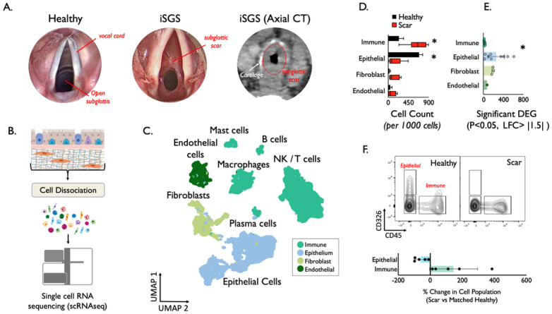
Background: Idiopathic subglottic stenosis (iSGS) is a rare fibrotic disease of the proximal airway affecting adult Caucasian women nearly exclusively. Life-threatening ventilatory obstruction occurs secondary to pernicious subglottic mucosal scar. Disease rarity and wide geographic patient distribution has previously limited substantive mechanistic investigation into iSGS pathogenesis.

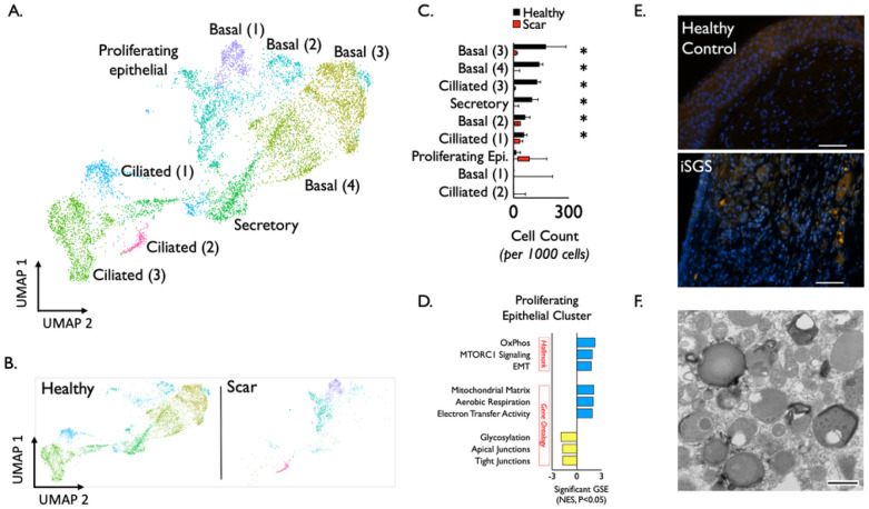
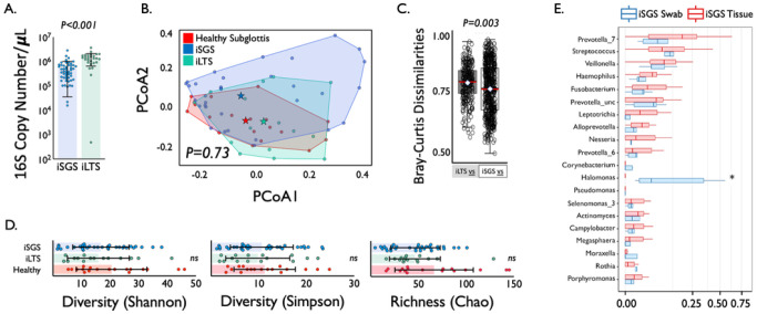
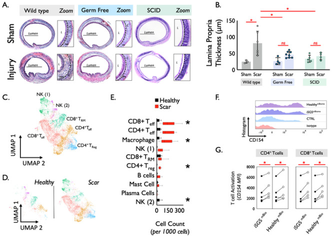
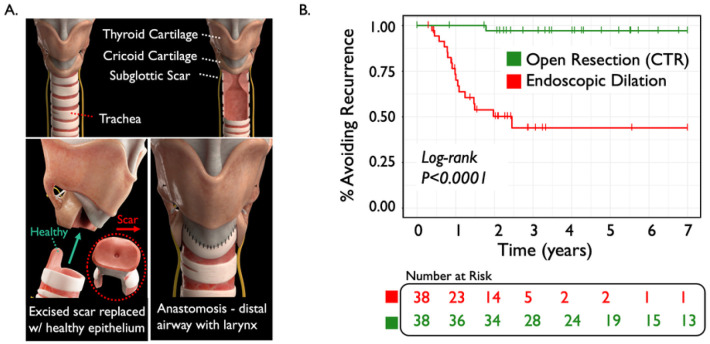
Result: By harnessing pathogenic mucosa from an international iSGS patient cohort and single-cell RNA sequencing, we unbiasedly characterize the cell subsets in the proximal airway scar and detail their molecular phenotypes. Results show that the airway epithelium in iSGS patients is depleted of basal progenitor cells, and the residual epithelial cells acquire a mesenchymal phenotype. Observed displacement of bacteria beneath the lamina propria provides functional support for the molecular evidence of epithelial dysfunction. Matched tissue microbiomes support displacement of the native microbiome into the lamina propria of iSGS patients rather than disrupted bacterial community structure. However, animal models confirm that bacteria are necessary for pathologic proximal airway fibrosis and suggest an equally essential role for host adaptive immunity. Human samples from iSGS airway scar demonstrate adaptive immune activation in response to the proximal airway microbiome of both matched iSGS patients and healthy controls. Clinical outcome data from iSGS patients suggests surgical extirpation of airway scar and reconstitution with unaffected tracheal mucosa halts the progressive fibrosis.

Conclusion: Our data support an iSGS disease model where epithelial alterations facilitate microbiome displacement, dysregulated immune activation, and localized fibrosis. These results refine our understanding of iSGS and implicate shared pathogenic mechanisms with distal airway fibrotic diseases.


Conflict of interest statement

COMPETING INTERESTS The authors of this manuscript declare no financial or other conflicts of interest to disclose as described by the journal Microbiome.

Figures

Figure 1
Figure 1
Anatomy of mucosal scar obstructing the subglottis in iSGS. Submucosal thickening with preserved cartilage seen on axial computed tomography (A). Workflow to generate single cell suspensions from subglottic scar (B). Uniform Manifold Approximation and Projection (UMAP) of jointly analyzed single-cell transcriptomes from 25,974 cells from 7 iSGS mucosal scar and 3 healthy mucosa annotated by cell type. (C) Cell types/states manually grouped into 4 broad tissue classes. (D) Quantification of cell types showed significantly increased immune cell populations in airway scar (p = 0.018) and significantly reduced epithelial cell numbers (p < 0.001) Boxes depict median and interquartile range wiskers show min to max, *P < 0.05 by Mann-Whitney U. (E) Number of differentially expressed genes (DEG) in each cell type in iSGS airway scar and paired healthy mucosa [negative binomial test, log fold change (FC) cutoff of 1.5 and adjusted P value of <0.05]. Representative flow cytometry of fresh single cell suspension from matched iSGS airway scar and healthy mucosa (n=5) confirming depletion of epithelial cells and increase in immune cells within iSGS airway scar when compared to matched healthy control. Graph depicting % change in cell population for matched scar/healthy mucosa for each of the five individual patients. Bar represents mean, error bars SEM, and dots show individual patients (F).
Figure 2
Figure 2
Detailed analysis of epithelial clusters (A) identified conserved transcriptional programs in basal (four clusters), ciliated (three clusters), secretory (one cluster), and a proliferating cell subset (one cluster) in both healthy and scar (B). Quantification of epithelial cell types/states show significant reduction in clusters comprising basal, secretory, and ciliated cells in scar samples (Boxes depict median and interquartile range, whiskers show min to max, *P < 0.05 by Mann-Whitney U) (C). In addition to the dramatic loss of basal cells within airway scar, geneset enrichment analysis for Hallmark genesets and Gene Ontology Biological Processes genesets demonstrate that residual proliferating epithelial cells express a molecular program for EMT. Blue represents genesets upregulated in scar epitheluim, yellow represents genesets down regulated in scar epithelium compared to healthy mucosa. FISH with the pan-bacterial probe Eub338 shows iSGS mucosa possessed signal for bacteria in the deeper lamina propria while healthy control did not. Scale bar represents 50μm (E). Transmission electron microscopy of separate iSGS scar specimen demonstrated numerous forms consistent with the size and shape of bacteria in the cell cytoplasm. Scale bar represents 500nm (F). mTORC1: mechanistic target of rapamycin complex 1, OxPhos: oxidative phosphorylation, EMT: epithelial-mesenchymal transition. FISH: Fluorescence in situ hybridization.
Figure 3
Figure 3
qPCR of 16S rRNA showed a significantly higher bacterial load (copy number per μL) in iLTS patients (green) compared to iSGS samples (blue) (P<0.0001) (A). Principal coordinate analysis of proximal microbiome in iSGS, post-intubation subglottic stenosis (iLTS), and healthy controls showing no significant differences in the centroids between the three groups (PerMANOVA adonis2 testing p = 0.06) (B). Binary comparisons between healthy and iLTS samples, and between heathy and iSGS samples using Bray-Curtis dissimilarities showed iSGS samples more closely resembled healthy controls (Wilcoxon rank sum test with continuity correction, p-value=0.001) (C). Microbial alpha diversity (Shannon and Simpson indicies), and richness (Chao1 index) shown for iLTS, iSGS, and healthy control samples (D). Individual sample values are represented as dots. Group mean depicted by box center line, and standard deviation represented by error bars. Microbial alpha diversity and richness were not significantly different between cases and controls. Comparison of bacterial abundance between sampling via mucosal swab vs. tissue biopsy in iSGS patients. The top 20 most abundant genera detected via the two methods with the genera abundance represented by boxplots; the median is represented by the center line of the box, and the interquartile range is represented by the upper and lower edges of the box. The vertical lines represent the whiskers (minimum and maximum, excluding outliers); outliers are represented as dots. Blue boxes are samples from Hillel et al., where the iSGS microbiome was sampled using swabs (N=5). Red boxes are samples from the tissue samples (N=37). Of the top 20 most abundant genera, the only significantly different genus between the two studies was Halomonas, which was relatively abundant in the iSGS swab samples but was not found in the iSGS tissue samples (E).
Figure 4
Figure 4
Established murine model of subglottic stenosis (A) demonstrated significant thickening of the lamina propria 14 days after epithelial injury in wild type mice (p=0.0036). However, no significant thickening of the lamina propria was observed in either germ free mice (p=0.56), or severe combined immunodeficient mice (SCID) (p=0.98) Wild type injury was significantly greater than both germ-free injury (p=0.025) and SCID injury (p=0.036) (B). Immune cell identification and functional characterization in iSGS Proximal Airway Scar via UMAP of jointly analyzed single-cell transcriptomes (C) Analysis of immune clusters identified conserved transcriptional programs in T cells (four clusters) and NK cells (two clusters), with differntial abundance in healthy mucosa and airway scar (D). Quantification of immune cell types in iSGS airway scar versus matched healthy mucosa. Boxes depict median and interquartile range wiskers show min to max, *P < 0.05 by Mann-Whitney U. (E). Single cell suspensions from 5 unique iSGS patients cultured in the presence of matched iSGS airway microbiome, the microbiome from an unrelated healthy subject, or left untreated. 24 hours after stimulation, expression levels of activation marker CD154 were quantified on CD4+ and CD8+ T cells. (F) Both the matched iSGS microbiome, as well as the microbiome from an unrelated healthy control significantly up-regulated CD154 when compared to untreated experimental controls (CD4+ matched iSGS microbiome vs untreated: p=0.007; CD4+ unrelated healthy microbiome vs untreated: p=0.04), (CD8+ matched iSGS microbiome vs untreated: p=0.005; CD8+ unrelated healthy microbiome vs untreated: p=0.03). No significant difference observed between cells treated with a matched iSGS microbiome or cells treated with an unrelated healthy microbiome (G).
Figure 5
Figure 5
Technique of open resection (cricotracheal resection: CTR) for idiopathic subglottic stenosis. Anatomic landmarks of the Thyroid and Cricoid cartilage along with the defined rings of the trachea. Obstructing mucosal scar arises below the vocal cords at the junction of the cricoid cartilage and proximal trachea (A). Oblique view indicates removal of affected proximal trachea as well as anterior arch of cricoid but conservation of posterolateral laminae and posterior cricoid to preserve recurrent laryngeal nerves (nerves indicated in yellow). The mucosal stenosis is excised, and a broad flap of healthy tracheal mucosa is pulled into place from below to resurface the airway (B). The continuity of the airway mucosa is resotred at the completion of the procedure (C). Kaplan-Meyer survival analysis showing patients undergoing CTR had a significantly lower rate of disease recurrence (Log-rank, P<0.0001) (D).
Figure 6
Figure 6
Proposed model of iSGS disease pathogenesis. Defects in epithelial barrier function allow translocation of the native microbial community deep into the airway mucosa and promote dysregulated immune activation leading to fibrotic remodeling and subsequent airway obstruction.

References

    1. Aldhahrani A., Powell J., Ladak S., Ali M., Ali S., Verdon B., Pearson J., and Ward C. (2018). The Potential Role of Bile Acids in Acquired Laryngotracheal Stenosis. Laryngoscope 128, 2029–2033. 10.1002/lary.27105. - DOI - PMC - PubMed
    1. Bachmaier K., Neu N., de la Maza L.M., Pal S., Hessel A., and Penninger J.M. (1999). Chlamydia infections and heart disease linked through antigenic mimicry. Science 283, 1335–1339. - PubMed
    1. Clark R.A. (2015). Resident memory T cells in human health and disease. Sci Transl Med 7, 269rv261. 10.1126/scitranslmed.3010641. - DOI - PMC - PubMed
    1. Daniero J.J., Ekbom D.C., Gelbard A., Akst L.M., and Hillel A.T. (2017). Inaugural Symposium on Advanced Surgical Techniques in Adult Airway Reconstruction: Proceedings of the North American Airway Collaborative (NoAAC). JAMA Otolaryngol Head Neck Surg 143, 609–613. 10.1001/jamaoto.2016.4126. - DOI - PMC - PubMed
    1. Drake V.E., Gelbard A., Sobriera N., Wohler E., Berry L.L., Hussain L.L., and Hillel A. (2020). Familial Aggregation in Idiopathic Subglottic Stenosis. Otolaryngol Head Neck Surg 163, 1011–1017. 10.1177/0194599820935402. - DOI - PMC - PubMed

Publication types