[Preprint]. 2023 May 24:2023.05.19.540713.
doi: 10.1101/2023.05.19.540713.

Disrupted memory T cell expansion in HIV-exposed uninfected infants is preceded by premature skewing of T cell receptor clonality

Affiliations

Disrupted memory T cell expansion in HIV-exposed uninfected infants is preceded by premature skewing of T cell receptor clonality

Sonwabile Dzanibe et al. bioRxiv. .

Update in

Abstract

While preventing vertical HIV transmission has been very successful, the increasing number of HIV-exposed uninfected infants (iHEU) experience an elevated risk to infections compared to HIV-unexposed and uninfected infants (iHUU). Immune developmental differences between iHEU and iHUU remains poorly understood and here we present a longitudinal multimodal analysis of infant immune ontogeny that highlights the impact of HIV/ARV exposure. Using mass cytometry, we show alterations and differences in the emergence of NK cell populations and T cell memory differentiation between iHEU and iHUU. Specific NK cells observed at birth were also predictive of acellular pertussis and rotavirus vaccine-induced IgG and IgA responses, respectively, at 3 and 9 months of life. T cell receptor Vβ clonotypic diversity was significantly and persistently lower in iHEU preceding the expansion of T cell memory. Our findings show that HIV/ARV exposure disrupts innate and adaptive immunity from birth which may underlie relative vulnerability to infections.

Keywords: HIV exposure; NK cels and antibody responses; T cell receptor; infant immunity.


Conflict of interest statement

Declaration of Interest All authors declare no conflict of interest of any of the material included in this manuscript.

Figures
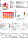
Figure 1:
Figure 1:
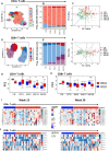
Immunophenotypic trajectory of infant peripheral blood mononuclear cells (PBMC) in the first 9 months of life. A) Dimensional reduction of infant PBMC Immune lineage clusters embedded on uniform manifold approximation and proximation (UMAP) with density preservation and table summarising immune cell cluster phenotype. B) Relative abundance of immune lineage clusters measured in cord blood (CB) and infant peripheral blood at birth (BTH), weeks (WK) 4, 15 and 36. C) Spearman’s rank correlation between proportion of immune lineage cell clusters and infants age from birth until week 36. D) Age related maturation trajectory of CD4+ and CD8+ T cells and NK cells depicted using multidimensional scaling coordinates derived from median marker expressions for each infant samples. E) Boxplots summarizing MDS coordinate associated with infant age for CD4+ and CD8+ T cells and NK cells.
Figure 2:
Figure 2:
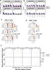
Divergent T cell memory differentiation in HIV-exposed uninfected infants (iHEU) compared to HIV-unexposed uninfected infants (iHUU). A & D) Uniform manifold approximation and proximation (UMAP) with density preservation showing dimensional reduction of FlowSOM clusters of CD4+ and CD8 T cells B &E) Relative abundances of CD4+ and CD8 + T cell clusters measured in cord blood (CB) and infant peripheral blood at birth (BTH), weeks (WK) 4, 15 and 36. C &F) Age related phenotypic composition of CD4+ and CD8+ T cells depicted as principal component (PC) coordinates of centred log-odd ratios of FlowSOM clusters for each infant samples and arrows indicating contribution of each cell cluster in scatter of PC components. G &H) Boxplots comparing PC coordinates of CD4+ and CD8+ T cells between iHEU and iHUU longitudinally. I & J) Generalized linear mixed model (GLMM) comparing the abundances of CD4+ and CD8+ T cell clusters between iHEU and iHUU at weeks 15 and 36.
Figure 3:
Figure 3:
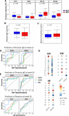
Early life immunophenotypic alteration in NK cells in HIV-exposed uninfected infants (iHEU) compared to HIV-unexposed uninfected infants (iHUU). A) Uniform manifold approximation and proximation (UMAP) with density preservation showing dimensional reduction of FlowSOM clusters on NK cells and table summarising immune cell cluster phenotype. B) Relative abundances of NK cell clusters measured in cord blood (CB) and infant peripheral blood at birth (BTH), weeks (WK) 4, 15 and 36. C) Age related phenotypic composition of NK cells depicted as principal component (PC) coordinates of centred log-odd ratios of FlowSOM clusters for each infant samples and arrows indicating contribution of each cell cluster in scatter of PC components. D) Boxplots comparing PC coordinates of NK cells between iHEU and iHUU
Figure 4:
Figure 4:
Premature CD4+ and CD8+ T cell receptor (TCR) repertoire skewing in HIV-exposed uninfected infants (iHEU) relative to HIV-unexposed uninfected infants (iHUU). A) Boxplots comparing Inverse Simpson TCR diversity scores between iHUU and iHEU at birth and weeks 4, 15 and 36. B) Boxplots comparing Chao1 TCR clonotype richness between iHUU and iHEU at birth and weeks 4, 15 and 36. C &D) Spearman’s rank correlation between naive CD4+ and CD8+ T cell Inverse Simpson scores and frequencies of FlowSOM clusters for CD4+ and CD8+ T cell clusters respectively. E). GLIPH analysis showing antigen specificity groups of the top 100 TCR clones that are significantly different between iHEU and iHUU and between naïve and memory CD4+ and CD8+ T cell subsets.
Figure 5
Figure 5
Association of vaccine antibody responses to immune cell phenotypes. A) Comparing IgG levels against pertussis between HIV-exposed uninfected infants (iHEU) and HIV-unexposed uninfected infants (iHUU), grey shaded area indicate threshold IgG levels for protective pertussis vaccine response. B & C) Boxplots comparing rotavirus specific IgA and neutralization titres between iHEU and iHUU at week 36. D, E & F) Summary of ROC analysis using the latent variable axis-1 derived from partial least square discriminate analysis (PLS-DA) of NK, CD4 and CD8 T cell clusters determined to be best predictors at birth and week 4 of pertussis antibody responses at weeks 15 and 36 and rotavirus antibody response at week 37. G) Spearman’s correlation between abundances FlowSOM clusters of CD4+ and CD8+ T cells and NK cells and anti-pertussis IgG titres measured at week 15 and 36.

References

    1. Perin J. et al. Global, regional, and national causes of under-5 mortality in 2000–19: an updated systematic analysis with implications for the Sustainable Development Goals. The Lancet Child and Adolescent Health 6, 106–115 (2022). - PMC - PubMed
    1. Zimmermann P. & Jones C. E. Factors That Influence Infant Immunity and Vaccine Responses. The Pediatric infectious disease journal 40, S40–S46 (2021). - PubMed
    1. Pasetti M. F. et al. Maternal determinants of infant immunity: Implications for effective immunization and maternal-child health. Vaccine 38, 4491–4494 (2020). - PMC - PubMed
    1. Sanidad K. Z. & Zeng M. Y. Neonatal gut microbiome and immunity. Current Opinion in Microbiology 56, 30–37 (2020). - PMC - PubMed
    1. Goenka A. & Kollmann T. R. Development of immunity in early life. Journal of Infection 71, S112–S120 (2015). - PubMed

Publication types