[Preprint]. 2023 May 18:2023.05.16.541010.
doi: 10.1101/2023.05.16.541010.

VirB, a key transcriptional regulator of Shigella virulence, requires a CTP ligand for its regulatory activities

Affiliations

VirB, a key transcriptional regulator of Shigella virulence, requires a CTP ligand for its regulatory activities

Taylor M Gerson et al. bioRxiv. .

Update in

Abstract

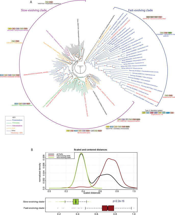
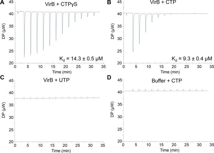
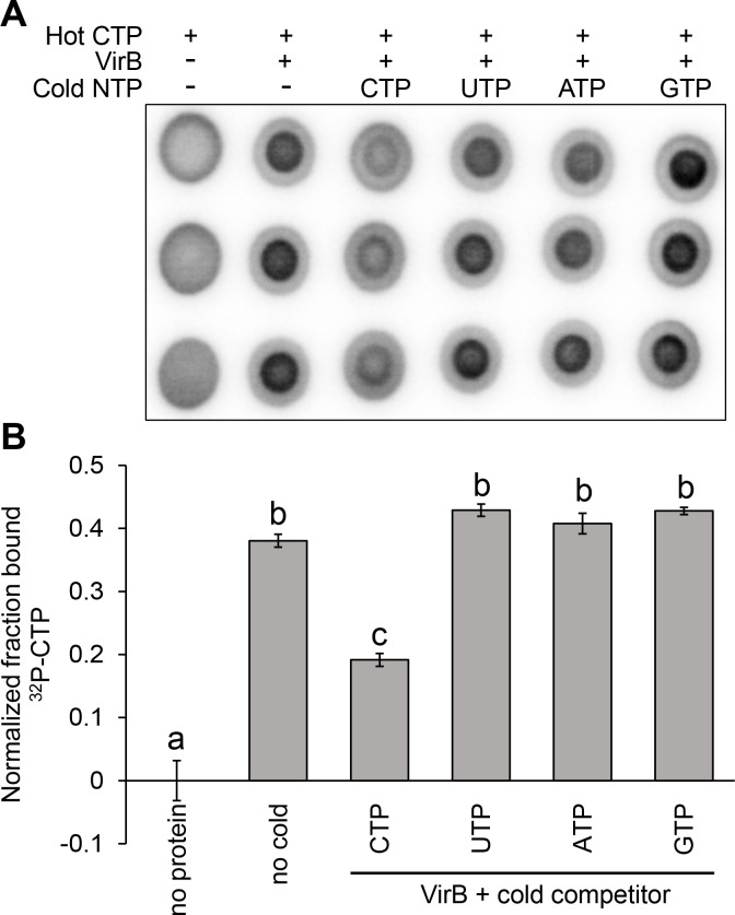
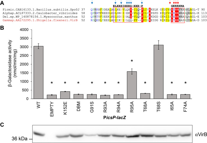
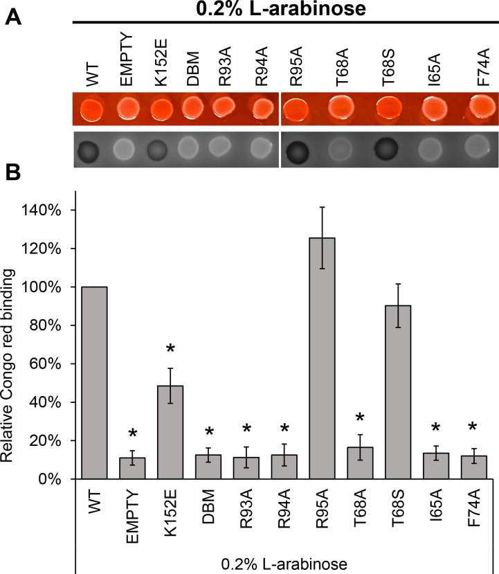
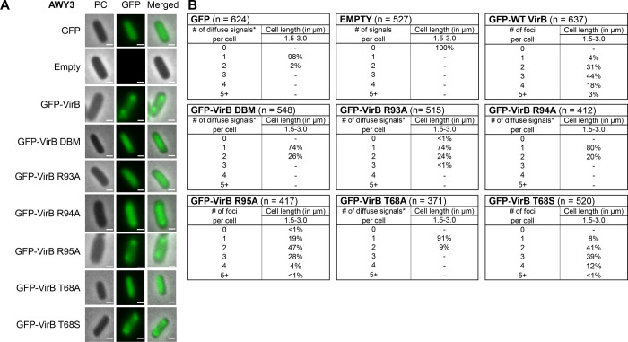
The VirB protein, encoded by the large virulence plasmid of Shigella spp., is a key transcriptional regulator of virulence genes. Without a functional virB gene, Shigella cells are avirulent. On the virulence plasmid, VirB functions to offset transcriptional silencing mediated by the nucleoid structuring protein, H-NS, which binds and sequesters AT-rich DNA, making it inaccessible for gene expression. Thus, gaining a mechanistic understanding of how VirB counters H-NS-mediated silencing is of considerable interest. VirB is unusual in that it does not resemble classic transcription factors. Instead, its closest relatives are found in the ParB superfamily, where the best-characterized members function in faithful DNA segregation before cell division. Here, we show that VirB is a fast-evolving member of this superfamily and report for the first time that the VirB protein binds a highly unusual ligand, CTP. VirB binds this nucleoside triphosphate preferentially and with specificity. Based on alignments with the best-characterized members of the ParB family, we identify amino acids of VirB likely to bind CTP. Substitutions in these residues disrupt several well-documented activities of VirB, including its anti-silencing activity at a VirB-dependent promoter, its role in generating a Congo red positive phenotype in Shigella , and the ability of the VirB protein to form foci in the bacterial cytoplasm when fused to GFP. Thus, this work is the first to show that VirB is a bona fide CTP-binding protein and links Shigella virulence phenotypes to the nucleoside triphosphate, CTP.

Importance: Shigella species cause bacillary dysentery (shigellosis), the second leading cause of diarrheal deaths worldwide. With growing antibiotic resistance, there is a pressing need to identify novel molecular drug targets. Shigella virulence phenotypes are controlled by the transcriptional regulator, VirB. We show that VirB belongs to a fast-evolving, primarily plasmid-borne clade of the ParB superfamily, which has diverged from versions that have a distinct cellular role - DNA partitioning. We are the first to report that, like classic members of the ParB family, VirB binds a highly unusual ligand, CTP. Mutants predicted to be defective in CTP binding are compromised in a variety of virulence attributes controlled by VirB. This study i) reveals that VirB binds CTP, ii) provides a link between VirB-CTP interactions and Shigella virulence phenotypes, and iii) broadens our understanding of the ParB superfamily, a group of bacterial proteins that play critical roles in many different bacteria.


Figures

Figure 1.
Figure 1.. Maximum likelihood tree of ParB protein sequences including the VirB-like fast-evolving sequences.
(A) Maximum likelihood tree of ParB protein sequences including the VirB-like fast-evolving sequences. The tree was computed using IQtree and bootstrapped with 1000 replicates. Nodes and branches corresponding to widely represented phylogenetic clades are colored according to the key. Protein sequences are denoted with their clade, gene and species name. The slow- and fast-evolving clades are marked and representative gene neighborhoods of distinct clades of the tree are shown. Genes in gene neighborhoods are shown as box arrows with the arrowhead pointing to the gene at the 3’ end. Clade abbreviations include: Actino: Actinomycetes, Alphap: Alpha-proteobacteria, Betapr: Beta-proteobacteria, candid: Dark-matter bacteria, Chloro: Chloroflexi, del.ep: Delta/epsilon proteobacteria, Firmic: Firmicutes, Gammap: Gamma-proteobacteria, Planct: Planctomycetes. (B) Differential evolutionary rates derived from pairwise centered and scaled maximum-likelihood distances shown as a kernel density and box plots.
Figure 2.
Figure 2.. The Shigella anti-silencing protein VirB binds a novel ligand, CTP.
ITC measurements with (A) 90 μM VirB-His6 and 3 mM CTPγS, (B) 45 μM VirB-His6 and 3 mM CTP, (C) 45 μM VirB-His6 and 3 mM UTP, and (D) VirB binding buffer and 3 mM CTP. In panels B and C, the VirB protein concentration was lowered to 45 μM to reach saturation and conserve protein.
Figure 3.
Figure 3.. VirB preferentially binds CTP over other NTP ligands found in the cell.
(A) DRaCALA images of competition assays assessing the ability of 500 μM indicated cold NTP to compete with binding interactions between 5 nM 32P-CTP and 50 μM VirB-His6. Ligand binding is indicated by a darker inner core staining due to rapid immobilization of the VirB-CTP complex. (B) Graph of normalized fraction bound (FB) calculated for each sample in Figure 3A. Assays were completed with three technical replicates and three biological replicates. Representative data are shown. Significance was calculated using a one-way ANOVA with post hoc Bonferroni, p < 0.05. Lowercase letters indicate statistical groups. Complete statistical analysis provided in Supplementary Table S3.
Figure 4.
Figure 4.. Key residues in the predicted CTP binding pocket are required for the anti-silencing activity of VirB.
(A) Multiple sequence alignment (MSA) of classic ParBs that bind CTP and VirB, generated using the Mafft program with a local pair algorithm. Full alignment and additional details provided in Figure S2. Taxonomic clade name, Genbank accession, and species name separated by dots are denoted . Consensus abbreviations and coloring scheme are as follows: hydrophobic (FWYILVACM), aromatic (FWY) and aliphatic (ILV) residues shaded yellow; charged (DEHKR) and basic (KRH) residues colored magenta, big (LIFMWYERKQ) residues shaded grey, alcohol-group (ST) residues colored red, polar (STECDRKHNQ) residues colored blue, small (AGSCDNPTV) residues colored green, and tiny (GAS) residues shaded green. Fully conserved residues are shaded red. Arrows indicate nucleotide-binding residues (blue) and catalytic residues (red). (B) β-Galactosidase assay used to assess the regulatory activity of pBAD-VirB derivatives at the VirB-dependent icsP promoter. Significance was calculated using a one-way ANOVA with post hoc Tukey HSD , p < 0.05.*, statistically significant compared to wild-type. Complete statistical analysis is provided in Supplementary Table S4. (C) Western Blot analysis using an anti-VirB antibody to assess protein production of pBAD-VirB mutants alongside a SeeBlue Plus2 Prestained Standard. Assays were completed with three biological replicates and repeated three times. Representative data are shown.
Figure 5.
Figure 5.. Key residues in the predicted CTP binding pocket are required for Shigella virulence phenotype.
(A) Congo red binding by S. flexneri virB::Tn5 harboring pBAD-VirB derivatives under inducing conditions. Images were captured using visible light (top) and blue light (Cy2) (bottom). (B) Quantitative analysis of Congo red binding S. flexneri virB::Tn5 harboring pBAD-VirB derivatives (induced). Relative Congo red binding was calculated as [(OD498/OD600)/(average (OD498/OD600)2457T pBAD)] × 100. Assays were completed with three biological replicates and repeated three times. Representative data are shown. Significance was calculated using a one-way ANOVA with post hoc Tukey HSD, p < 0.05. *, statistically significant compared to wild-type. Complete statistical analysis is provided in Supplementary Table S5
Figure 6.
Figure 6.. Key residues in the CTP binding pocket are required for VirB focus formation in vivo.
(A) Live-cell imaging of GFP-VirB derivatives in a virB mutant strain of S. flexneri (AWY3) induced with 0.02% L-arabinose. Phase-contrast, PC, (left column), fluorescence (middle column) (GFP row, 28-ms exposure; GFP-VirB derivatives and empty rows, 98-ms exposure), and merged (right column). Bars represent 1 μM. (B) Quantification of fluorescent signals observed during live-cell imaging of GFP-VirB derivatives using MicrobeJ. Within tables, a hyphen indicates that no cells were found in this category in any of the images captured; *, maxima detected by MicrobeJ. Complete statistical analysis is provided in Supplementary Table S6 and field-of-view images are found in Figure S5.

Similar articles

References

    1. Hawkins HP. 1909. The identity of British ulcerative colitis and tropical bacillary dysentery. Br Med J 2:1331–2. - PMC - PubMed
    1. Labrec EH, Schneider H, Magnani TJ, Formal SB. 1964. Epithelial cell penetration as an essential step in the pathogenesis of bacillary dysentery. J Bacteriol 88:1503–18. - PMC - PubMed
    1. Niyogi SK. 2007. Increasing antimicrobial resistance--an emerging problem in the treatment of shigellosis. Clin Microbiol Infect 13:1141–3. - PubMed
    1. Niyogi SK. 2005. Shigellosis. J Microbiol 43:133–43. - PubMed
    1. Tobe T, Nagai S, Okada N, Adler B, Yoshikawa M, Sasakawa C. 1991. Temperature-regulated expression of invasion genes in Shigella flexneri is controlled through the transcriptional activation of the virB gene on the large plasmid. Mol Microbiol 5:887–93. - PubMed

Publication types