[Preprint]. 2023 Jan 24:2023.01.24.525378.
doi: 10.1101/2023.01.24.525378.

Mast cell-derived BH4 is a critical mediator of postoperative pain

Affiliations

Mast cell-derived BH4 is a critical mediator of postoperative pain

Philipp Starkl et al. bioRxiv. .

Update in

  • Mast cell-derived BH4 and serotonin are critical mediators of postoperative pain.
    Starkl P, Jonsson G, Artner T, Turnes BL, Gail LM, Oliveira T, Jain A, Serhan N, Stejskal K, Lakovits K, Hladik A, An M, Channon KM, Kim H, Köcher T, Weninger W, Stary G, Knapp S, Klang V, Gaudenzio N, Woolf CJ, Tikoo S, Jain R, Penninger JM, Cronin SJF. Starkl P, et al. Sci Immunol. 2024 Aug 23;9(98):eadh0545. doi: 10.1126/sciimmunol.adh0545. Epub 2024 Aug 23. Sci Immunol. 2024. PMID: 39178277

Abstract

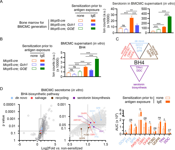
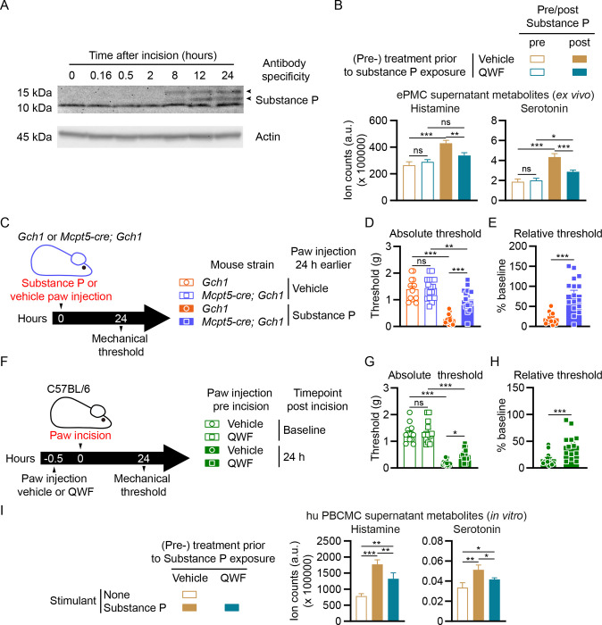
Postoperative pain affects most patients after major surgery and can transition to chronic pain. Here, we discovered that postoperative pain hypersensitivity correlated with markedly increased local levels of the metabolite BH4. Gene transcription and reporter mouse analyses after skin injury identified neutrophils, macrophages and mast cells as primary postoperative sources of GTP cyclohydrolase-1 (Gch1) expression, the rate-limiting enzyme in BH4 production. While specific Gch1 deficiency in neutrophils or macrophages had no effect, mice deficient in mast cells or mast cell-specific Gch1 showed drastically decreased postoperative pain after surgery. Skin injury induced the nociceptive neuropeptide substance P, which directly triggers the release of BH4-dependent serotonin in mouse and human mast cells. Substance P receptor blockade substantially ameliorated postoperative pain. Our findings underline the unique position of mast cells at the neuro-immune interface and highlight substance P-driven mast cell BH4 production as promising therapeutic targets for the treatment of postoperative pain.


Conflict of interest statement

Declaration of Interests The authors declare no competing financial interests.

Figures

Figure 1.
Figure 1.. Tissue damage and pain-like hypersensitivity is associated with Gch1-expressing immune cells in the incision-injured paw.
(A) Experimental scheme for (B to D). C57BL/6 wild type mice were subjected to paw incision. Contralateral (Contra) and ipsilateral (Ipsi) paw mechanical pain sensitivity and paw BH4 levels were assessed at the indicated time points after incision. (B) Time course of mechanical pain sensitivity relative to baseline at the indicated time points after incision (left panel; n=28) and thermal pain sensitivity 24 and 48 hours after incision compared to baseline before incision (right panel; n=10). (C) Thickness change (compared to baseline) of Contra and Ipsi paws 24 hours after incision (n=10–21). (D) BH4 levels in Contra and Ipsi paws 24 hours after incision (n=26). (E) Experimental scheme and measurement of BH4 levels in the Contra and Ipsi L3-L4 dorsal root ganglion (DRG) tissues of incision injury- (n=4) wild type mice and a spared nerve injury (SNI)-treated (n=6) wild type mouse as control. (F) Experimental scheme and representative immunofluorescence (immunofl.) images of anti-beta-TUBULIN-III (ßTUB3) and anti-GFP (Gch1-GFP) staining in the Ipsi L3-L4 DRG tissues of incision injury-(representative of n=3) and spared nerve injury (SNI)-treated (representative of n=3) Gch1-GFP reporter mice. Arrows indicate Gch1-GFP-positive cells. Scale bars represent 50 μm. (G) Experimental scheme for (H and I). Gch1flox/flox (Gch1) control mice or Gch1 mice expressing tamoxifen-inducible cre recombinase in sensory neurons (Brn3A-creERT; Gch1flox/flox; Brn3A-creERT; Gch1) mice were treated by daily intraperitoneal tamoxifen injections (2mg/mouse) 28 – 24 days before paw incision. Contra and Ipsi paw mechanical pain sensitivity and paw BH4 levels were assessed at the indicated time points after incision. (H) Absolute mechanical threshold at baseline and relative (to baseline) mechanical threshold kinetics (n=6). (I) Paw BH4 levels (n=3). (B to D, H) Mann-Whitney test; (E, I) One-Way ANOVA with Tukey’s multiple comparisons test; (H) Two-Way ANOVA with Sidak’s multiple comparisons test (comparing individual time points) and Two-Way ANOVA with repeated measures with Geisser-Greenhouse correction (overall comparison); * P ≤ 0.05, ** P ≤ 0.01, *** P ≤ 0.001, ns – not significant. Error bars indicate (B to E) SEM or (H and I) SD. Symbols in bar graphs represent individual mice.
Figure 2.
Figure 2.. Myeloid immune cells express Gch1 in injured skin.
(A) Mouse Gch1 gene expression levels in immune cell populations (n=1–4) derived from the ImmGen Microarray dataset using the Gene Expression Commons platform (Table S1). (B and C) Twenty-four hours after incision, skin cells from Contra and Ipsi paws of Gch1-GFP reporter (one pool of 5) or wild type C57BL/6 (one pool of 5) mice were analyzed by flow cytometry. (B) Left panel: experimental scheme; middle panel: histograms depicting Gch1-GFP signal of live CD45+ cells; right panel: Gch1-GFP mean fluorescence intensity (MFI) signals of live CD45+ cells. (C) Representation of immune cell populations meeting threshold criteria (see methods and Fig. S1), depicting Gch1-GFP MFI (y-axis) and % among GFP+ leukocytes (x-axis). (D) Experimental scheme for (E and F). Control Gch1flox/flox (Gch1) mice and animals with neutrophil-specific Gch1 deficiency (MRP8-cre; Gch1) were subjected to incision injury. Contralateral (Contra) and ipsilateral (Ipsi) paw mechanical pain sensitivity and BH4 levels were assessed 24 hours after incision. (E) Absolute mechanical threshold at baseline and relative (to baseline) mechanical threshold kinetics (n=7). (F) Paw BH4 levels (n=4–6). (G) Experimental scheme for (H and I). Gch1flox/flox (Gch1) mice or Gch1 mice expressing cre recombinase under control of the lysozyme promoter in macrophages (as well as in proportions of neutrophils and monocytes; LyzM-cre; Gch1) mice were treated by paw incision. Contra and Ipsi paw mechanical pain sensitivity and paw BH4 levels were assessed at indicated time points after incision. (H) Absolute mechanical threshold at baseline and relative (to baseline) mechanical threshold kinetics (n=10). (I) Paw BH4 levels (n=5). (F, I) One-Way ANOVA with Tukey’s multiple comparisons test; * P ≤ 0.05, ** P ≤ 0.01, *** P ≤ 0.001, ns – not significant. Error bars indicate SD. Symbols in bar graphs represent individual mice.
Figure 3.
Figure 3.. Mast cells are major contributors to postoperative pain-like hypersensitivity.
(A) Representative skin immunofluorescence images of sulforhodamin-101-labelled avidin (AvidinSR) and anti-GFP staining in Ipsi paws of wild type and Gch1-GFP reporter mice 24 hours post incision injury. Scale bars represent 20 μm. (B) Experimental scheme and measurements of serotonin and histamine levels in Contra and Ipsi paws 24 hours after incision injury in wild type animals (n=30–33). (C) Experimental scheme for (D and E). Postoperative pain kinetics and paw metabolites were assessed in control (Mcpt5-cre) and mast cell-depleted (Mcpt5-cre; DTA) mice. (D) Absolute mechanical threshold at baseline and relative (to baseline) mechanical threshold kinetics (n=6). (E) Paw tissue metabolites 24 hours post incision injury of Contra and Ipsi paws (n=5–6). (B) Mann-Whitney test; (E) One-Way ANOVA with Tukey’s multiple comparisons test; (D) Two-Way ANOVA with Sidak’s multiple comparisons test (comparing individual time points) and Two-Way ANOVA with repeated measures with Geisser-Greenhouse correction (overall comparison); * P ≤ 0.05, ** P ≤ 0.01, *** P ≤ 0.001, ns – not significant. Error bars indicate (B, D) SEM or (E) SD.
Figure 4.
Figure 4.. Mast cell-specific Gch1 deletion and overexpression modulates postoperative mechanical hypersensitivity.
(A) Experimental scheme for (B – E). Postoperative mechanical and thermal pain hypersensitivity as well as paw thickness and metabolites were assessed in control (Gch1) and Mcpt5-cre; Gch1 mice. (B) Absolute mechanical threshold at baseline and relative (to baseline) mechanical threshold kinetics (n=11–14). (C) Thermal pain sensitivity 24 hours after incision compared to baseline before incision (n=5–6). (D) Paw tissue metabolites of Contra and Ipsi paws 24 hours post incision injury (n=4–12). (E) Contra and Ipsi paw thickness change 24 hours post incision injury relative to baseline (before incision) (n=5–7). (F) Experimental scheme for (G and H). Postoperative pain kinetics and metabolites in contralateral (Contra) and ipsilateral (Ipsi) paws in control (GOE) and Mcpt5-cre; GOE mice. (G) Absolute mechanical threshold at baseline and relative (to baseline) mechanical threshold kinetics (n=6–8). (H) Contra and Ipsi paw tissue metabolites 24 hours post incision injury (n=3–4). (B, G) Two-Way ANOVA with Sidak’s multiple comparisons test (comparing individual time points) and Two-Way ANOVA with repeated measures with Geisser-Greenhouse correction (overall comparison); (C) Mann-Whitney test; (D, E, H) One-Way ANOVA with Tukey’s multiple comparisons test; * P ≤ 0.05, ** P ≤ 0.01, *** P ≤ 0.001, ns – not significant. Error bars indicate (B, D [BH4 and serotonin panels], E, G) SEM or (C, D [histamine panel], H) SD. Symbols in bar graphs represent individual mice.
Figure 5.
Figure 5.. Activated mast cells release BH4, serotonin as well as their biosynthetic enzymes upon degranulation.
(A, B) Bone marrow-derived cultured mast cells (BMCMCs) were generated from control (Mcpt5-cre) or Mcpt5-cre; Gch1, or Mcpt5-cre; GOE mice and activated by IgE and antigen for 1 hour, followed by analysis of (A) serotonin or (B) BH4 levels in the supernatant. (C) Schematic depicting the de novo, salvage and recycling arms of the BH4 pathway as well as the serotonin synthesis pathway. GTP, guanosine triphosphate; GCH1, GTP cyclohydrolase I; PTS, 6-Pyruvoyl tetrahydropterin synthase; SPR, sepiapterin reductase; AKR1, aldo-keto reductase family 1; CBR, carbonyl reductase family; DHFR, dihydrofolate reductase; QDPR, quinoid dihydropteridine reductase; PCDB, pterin-4alpha-carbinolamine dehydratase; TPH, tryptophan hydroxylase; DDC, dopa decarboxylase. (D; left and middle panel) Volcano blots of mass spectrometry results representing fold change (x axis) and statistical significance (y axis) of detected proteins in supernatant of IgE-sensitized vs. non-sensitized mast cells after 1 hour antigen exposure (the middle panel is a magnification of the data range indicated by the rectangle in the left panel). Circle colors in the left and middle panels indicate an association with the respective BH4 or serotonin biosynthesis pathways (respective proteins are labelled in the middle panel) as shown in (C) and Figure S5D. The right panel depicts the absolute quantification (area under the curve – AUC) of selected BH4 or serotonin biosynthesis pathways components detected by mass spectrometry. Dotted lines indicated where p=0.05 (horizontal line on the y axis) and a fold change of 2 (vertical line on the x axis). (A, B) One-Way ANOVA with Tukey’s multiple comparisons test; (D, middle and right panels) t-test; * P ≤ 0.05, ** P ≤ 0.01, *** P ≤ 0.001, ns – not significant. Error bars indicate SD. Circular symbols in volcano blots represent detected proteins.
Figure 6.
Figure 6.. Interference with Substance P-mediated mast cell degranulation alleviates postoperative mechanical hypersensitivity.
(A) Western blots of Substance P (upper picture) and actin (lower picture; loading control) in the skin tissue of paws from wild type mice collected before or sampled at the indicated time points after incision. Arrowheads indicate positions of α- and β-preprotachykinin. (B) Enriched peritoneal mast cells (ePMCs) of C57BL/6 wild type mice were pre-treated with vehicle or 100 μM QWF prior to stimulation. Histamine and serotonin in supernatants were analyzed after 1 hour stimulation with 10 μM Substance P. (C) Experimental scheme for (D and E). Control (Gch1) and Mcpt5-cre; Gch1 received injections of either vehicle (saline) or Substance P (50 μg) into the left hind paw. Paw mechanical thresholds were assessed 24 hours after injection. (D) Mechanical thresholds 24 hours post injection (n=14–16). (E) Mechanical thresholds relative to vehicle 24 hours post injection (n=14–16). (F) Experimental scheme for (G and H). C57BL/6 wild type mice were injected with either vehicle or QWF (20 μl of a 0.5mM solution) into the left hind paw 30 minutes before baseline mechanical threshold assessment, followed by paw incision and 24 hours later by mechanical threshold assessment. (G) Absolute mechanical thresholds before and 24 hours post incision (n=13). (H) Mechanical thresholds relative to baseline 24 hours post incision (n=13). (I) Human peripheral blood-derived cultured mast cells (hu PBCMCs) were pre-treated with vehicle or 100 μM QWF prior to simulation (or left unstimulated). Histamine and serotonin in supernatants were analyzed after 1 hour incubation with (or without) 100 μM Substance P. (B, D, G, I) One-Way ANOVA with Tukey’s multiple comparisons test; (E, H) Mann-Whitney test; * P ≤ 0.05, ** P ≤ 0.01, *** P ≤ 0.001, ns – not significant. Error bars indicate (B, I) SD or (D, E, G, H) SEM. Symbols in bar graphs represent individual mice.

References

    1. Abram C.L., Roberge G.L., Hu Y., and Lowell C.A.. 2014. Comparative analysis of the efficiency and specificity of myeloid-Cre deleting strains using ROSA-EYFP reporter mice. J Immunol Methods 408:89–100. - PMC - PubMed
    1. Aguilera-Lizarraga J., Florens M.V., Viola M.F., Jain P., Decraecker L., Appeltans I., Cuende-Estevez M., Fabre N., Van Beek K., Perna E., Balemans D., Stakenborg N., Theofanous S., Bosmans G., Mondelaers S.U., Matteoli G., Ibiza Martinez S., Lopez-Lopez C., Jaramillo-Polanco J., Talavera K., Alpizar Y.A., Feyerabend T.B., Rodewald H.R., Farre R., Redegeld F.A., Si J., Raes J., Breynaert C., Schrijvers R., Bosteels C., Lambrecht B.N., Boyd S.D., Hoh R.A., Cabooter D., Nelis M., Augustijns P., Hendrix S., Strid J., Bisschops R., Reed D.E., Vanner S.J., Denadai-Souza A., Wouters M.M., and Boeckxstaens G.E.. 2021. Local immune response to food antigens drives meal-induced abdominal pain. Nature 590:151–156. - PMC - PubMed
    1. Akula S., Paivandy A., Fu Z., Thorpe M., Pejler G., and Hellman L.. 2020. How Relevant Are Bone Marrow-Derived Mast Cells (BMMCs) as Models for Tissue Mast Cells? A Comparative Transcriptome Analysis of BMMCs and Peritoneal Mast Cells. Cells 9: - PMC - PubMed
    1. Aley K.O., McCarter G., and Levine J.D.. 1998. Nitric oxide signaling in pain and nociceptor sensitization in the rat. J Neurosci 18:7008–7014. - PMC - PubMed
    1. Arai H., Takahashi R., Sakamoto Y., Kitano T., Mashita O., Hara S., Yoshikawa S., Kawasaki K., and Ichinose H.. 2020. Peripheral tetrahydrobiopterin is involved in the pathogenesis of mechanical hypersensitivity in a rodent postsurgical pain model. Pain 161:2520–2531. - PubMed

Publication types