

A novel therapeutic approach for endometriosis using adipose-derived stem cell-derived conditioned medium- A new hope for endometriotic patients in improving fertility
- PMID: 37293500
- PMCID: PMC10244723
- DOI: 10.3389/fendo.2023.1158527
A novel therapeutic approach for endometriosis using adipose-derived stem cell-derived conditioned medium- A new hope for endometriotic patients in improving fertility
Abstract
Introduction: Endometriosis is defined as the growth of endometrial glands and stromal cells in a heterotopic location with immune dysregulation. It usually leads to chronic pelvic pain and subfertility. Although various treatments are available, the recurrence rate remains high. Adipose tissue is an abundant source of multipotent mesenchymal adipose-derived stem cells (ADSCs). ADSCs display effects on not only tissue regeneration, but also immune regulation. Thus, the current study aims to test the effects of ADSCs on the growth of endometriosis.
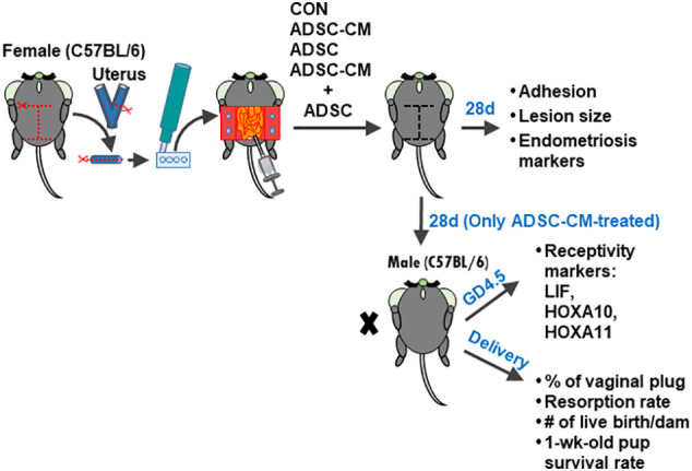
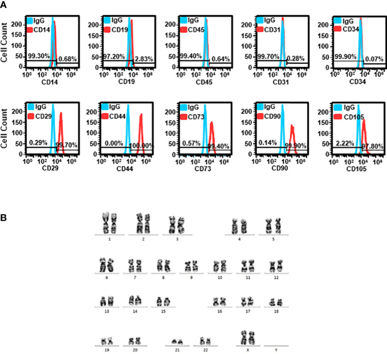
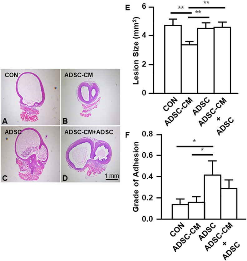
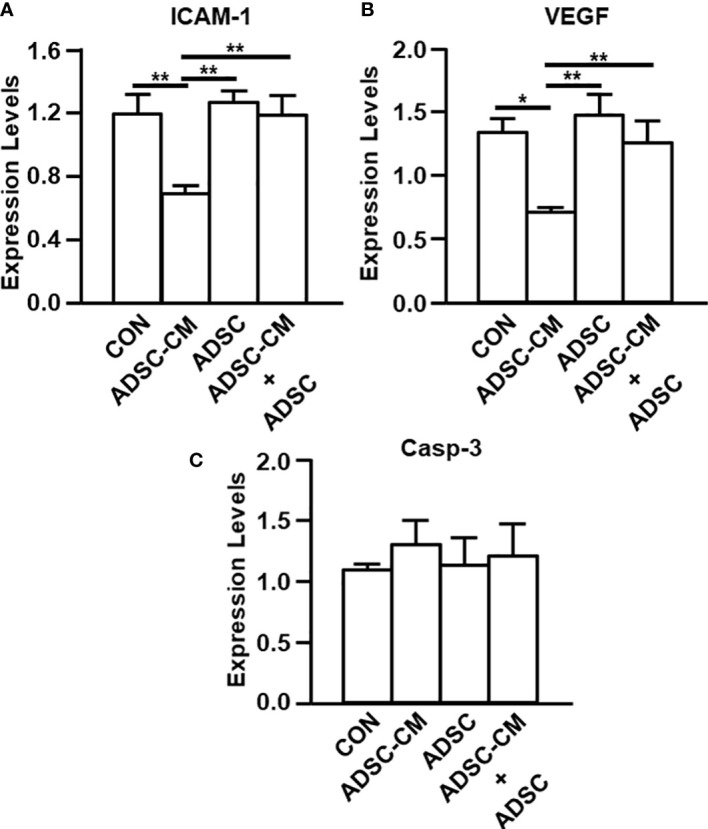
Methods: ADSCs isolated from lipoaspiration-generated adipose tissue and their conditioned medium (ADSC-CM) were subjected to quality validation, including karyotyping as well as growth promotion and sterility tests for microbial contamination under Good Tissue Practice and Good Manufacturing Practice regulations. An autologous endometriosis mouse model was established by suturing endometrial tissue to peritoneal wall followed by treating with DMEM/F12 medium, ADSC-CM, ADSCs or ADSC-CM+ADSCs for 28 days. The area of endometriotic cysts and the degree of pelvic adhesion were measured. ICAM-1, VEGF and caspase 3 expression was assessed by quantitative reverse transcription polymerase chain reaction (qRT-PCR) and immunohistochemistry. Moreover, the mice were allowed to mate and deliver. The pregnancy outcomes were recorded. The ADSC-CM was subjected to proteomics analysis with further data mining with Ingenuity Pathway Analysis (IPA).
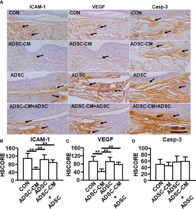
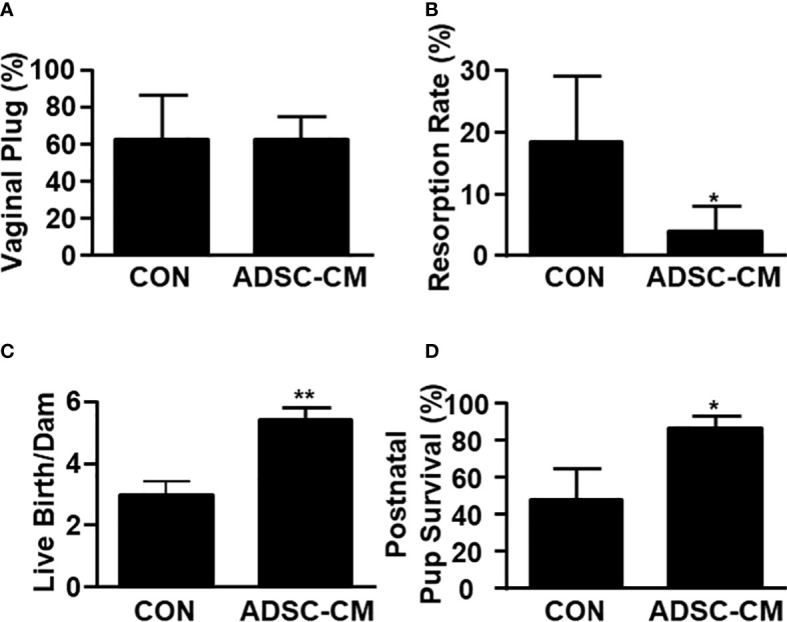
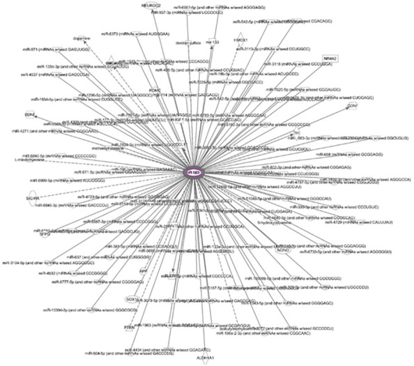
Results: Both ADSC-CM and ADSCs passed quality validation. ADSC-CM reduced the area of endometriotic cysts. The inhibition by ADSC-CM was obliterated by adding ADSCs. The presence of ADSCs with or without ADSC-CM increased the peritoneal adhesion. ADSC-CM inhibited ICAM-1 and VEGF mRNA and protein expression, whereas the addition of ADSCs not only did not inhibit by itself, but also blocked the inhibition by ADSC-CM. The resorption rate was reduced by ADSC-CM. The number of live birth/dam and the survival rate of pup at 1 week-old were both increased by ADSC-CM in mice with endometriosis. IPA demonstrated that PTX3 was potentially critical for the inhibition of endometriosis by ADSC-CM due to its anti-inflammatory and antiangiogenic properties as well as its importance in implantation.
Conclusion: ADSC-CM inhibited endometriosis development and improved pregnancy outcomes in mice. Potential translation to clinical treatment for human endometriosis is expected.
Keywords: PTX3; adipose-derived stem cell; conditioned medium; endometriosis; proteomics.
Copyright © 2023 Huang, Huang, Huang, Cheng, Yu, Lai, Hung, Chang and Shiu.
Conflict of interest statement
The authors declare that the research was conducted in the absence of any commercial or financial relationships that could be construed as a potential conflict of interest.
Figures

Similar articles
-
[Effects of hypoxia-pretreated rat adipose-derived mesenchymal stem cells conditioned medium on wound healing of rats with full-thickness defects].Zhonghua Shao Shang Za Zhi. 2020 Sep 20;36(9):803-812. doi: 10.3760/cma.j.cn501120-20200508-00258. Zhonghua Shao Shang Za Zhi. 2020. PMID: 32972065 Chinese.
-
[Effects of mouse adipose-derived stem cell conditioned medium on the apoptosis of keratinocytes induced by thermal injury in vitro].Zhonghua Shao Shang Za Zhi. 2014 Apr;30(2):102-8. Zhonghua Shao Shang Za Zhi. 2014. PMID: 24989653 Chinese.
-
Conditioned Medium from Adipose-Derived Stem Cells (ADSCs) Promotes Epithelial-to-Mesenchymal-Like Transition (EMT-Like) in Glioma Cells In vitro.Mol Neurobiol. 2016 Dec;53(10):7184-7199. doi: 10.1007/s12035-015-9585-4. Epub 2015 Dec 19. Mol Neurobiol. 2016. PMID: 26687184
-
The Basic Mechanism of Hair Growth Stimulation by Adipose-derived Stem Cells and Their Secretory Factors.Curr Stem Cell Res Ther. 2017;12(7):535-543. doi: 10.2174/1574888X12666170829161058. Curr Stem Cell Res Ther. 2017. PMID: 28875863 Review.
-
Therapeutic applications of adipose cell-free derivatives: a review.Stem Cell Res Ther. 2020 Jul 22;11(1):312. doi: 10.1186/s13287-020-01831-3. Stem Cell Res Ther. 2020. PMID: 32698868 Free PMC article. Review.
Cited by
-
Medical management of endometriosis.Curr Opin Obstet Gynecol. 2024 Oct 1;36(5):353-361. doi: 10.1097/GCO.0000000000000983. Epub 2024 Aug 17. Curr Opin Obstet Gynecol. 2024. PMID: 39159261 Free PMC article. Review.
-
Stem cell treatments for female reproductive disorders: a comprehensive review.J Ovarian Res. 2025 Jul 24;18(1):161. doi: 10.1186/s13048-025-01750-y. J Ovarian Res. 2025. PMID: 40708035 Free PMC article. Review.
References
Publication types
MeSH terms
Substances
LinkOut - more resources
Full Text Sources
Medical
Research Materials
Miscellaneous