

Kinetically inert manganese (II)-based hybrid micellar complexes for magnetic resonance imaging of lymph node metastasis
- PMID: 37293571
- PMCID: PMC10244211
- DOI: 10.1093/rb/rbad053
Kinetically inert manganese (II)-based hybrid micellar complexes for magnetic resonance imaging of lymph node metastasis
Abstract
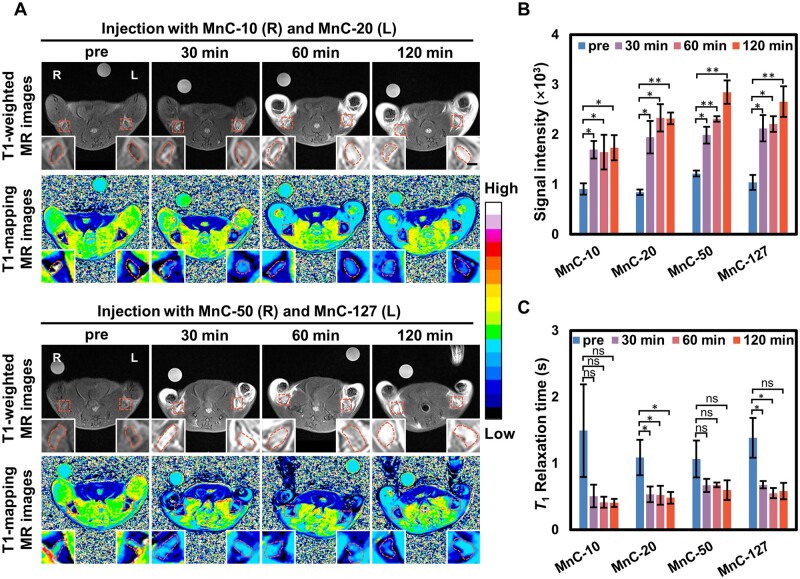
The localization and differential diagnosis of the sentinel lymph nodes (SLNs) are particularly important for tumor staging, surgical planning and prognosis. In this work, kinetically inert manganese (II)-based hybrid micellar complexes (MnCs) for magnetic resonance imaging (MRI) were developed using an amphiphilic manganese-based chelate (C18-PhDTA-Mn) with reliable kinetic stability and self-assembled with a series of amphiphilic PEG-C18 polymers of different molecular weights (C18En, n = 10, 20, 50). Among them, the probes composed by 1:10 mass ratio of manganese chelate/C18En had slightly different hydrodynamic particle sizes with similar surface charges as well as considerable relaxivities (∼13 mM-1 s-1 at 1.5 T). In vivo lymph node imaging in mice revealed that the MnC MnC-20 formed by C18E20 with C18-PhDTA-Mn at a hydrodynamic particle size of 5.5 nm had significant signal intensity brightening effect and shortened T1 relaxation time. At an imaging probe dosage of 125 μg Mn/kg, lymph nodes still had significant signal enhancement in 2 h, while there is no obvious signal intensity alteration in non-lymphoid regions. In 4T1 tumor metastatic mice model, SLNs showed less signal enhancement and smaller T1 relaxation time variation at 30 min post-injection, when compared with normal lymph nodes. This was favorable to differentiate normal lymph nodes from SLN under a 3.0-T clinical MRI scanner. In conclusion, the strategy of developing manganese-based MR nanoprobes was useful in lymph node imaging.
Keywords: T1 contrast agent; amphiphilic manganese chelate; kinetically inert; magnetic resonance imaging; sentinel lymph node.
© The Author(s) 2023. Published by Oxford University Press.
Figures


References
-
- Fujimoto Y, Okuhata Y, Tyngi S, Namba Y, Oku N.. Magnetic resonance lymphography of profundus lymph nodes with liposomal gadolinium-diethylenetriamine pentaacetic acid. Biol Pharm Bull 2000;23:97–100. - PubMed
-
- Schütz G, Lohrke J, Pietsch H.. Lymph node staging using dedicated magnetic resonance contrast agents—the accumulation mechanism revisited. Wiley Interdiscip Rev Nanomed Nanobiotechnol 2015;7:238–49. - PubMed
-
- Lu B, Wang H, Lu Q, Tang Z, Dou H, Dai T, Li S.. Novel hybrid dextran-gadolinium nanoparticles as high-relaxivity T1 magnetic resonance imaging contrast agent for mapping the sentinel lymph node. J Comput Assist Tomogr 2019;43:350–7. - PubMed
-
- Fu X, Fu S, Cai Z, Jin R, Xia C, Lui S, Song B, Gong Q, Ai H.. Manganese porphyrin/ICG nanoparticles as magnetic resonance/fluorescent dual-mode probes for imaging of sentinel lymph node metastasis. J Mater Chem B 2022;10:10065–74. - PubMed
-
- Han M, Kang R, Zhang C.. Lymph node mapping for tumor micrometastasis. ACS Biomater Sci Eng 2022;8:2307–20. - PubMed
LinkOut - more resources
Full Text Sources
