Case Reports
. 2023 Jun 7;10(2):78-85.
doi: 10.22551/2023.39.1002.10246. eCollection 2023.

COVID-19 related acute necrotizing encephalopathy presenting in the early postoperative period

Affiliations
Case Reports

COVID-19 related acute necrotizing encephalopathy presenting in the early postoperative period

Elissavet Symeonidou et al. Arch Clin Cases. .

Abstract

Besides respiratory and gastrointestinal symptoms, SARS-CoV-2 also has potential neurotropic effects. Acute hemorrhagic necrotizing encephalopathy is a rare complication of Covid-19. This article presents a case of an 81-year-old female, fully vaccinated, who underwent laparoscopic transhiatal esophagectomy due to gastroesophageal junction cancer. In the early postoperative period, the patient developed persistent fever accompanied by acute quadriplegia, impaired consciousness, and no signs of respiratory distress. Imaging with Computed Tomography and Magnetic Resonance revealed multiple bilateral lesions both in gray and white matter, as well as pulmonary embolism. Covid-19 infection was added to the differential diagnosis three weeks later, after other possible causes were excluded. The molecular test obtained at that time for coronavirus was negative. However, the high clinical suspicion index led to Covid-19 antibody testing (IgG and IgA), which confirmed the diagnosis. The patient was treated with corticosteroids with noticeable clinical improvement. She was discharged to a rehabilitation center. Six months later, the patient was in good general condition, although a neurological deficit was still present. This case indicates the significance of a high clinical suspicion index, based on a combination of clinical manifestations and neuroimaging, and the confirmation of the diagnosis with molecular and antibody testing. Constant awareness of a possible Covid-19 infection among hospitalized patients is mandatory.

Keywords: acute hemorrhagic necrotizing encephalopathy; case report; neuroimaging diagnosis; neurologic manifestations, covid-19; prompt treatment.


Figures

Fig. 1
Fig. 1
Magnetic Resonance Imaging immediately after the acute onset of quadrivbplegia: a, b: T2/FLAIR sequence, axial view, multiple lesions of high MR signal located in white matter, more intense in bilateral frontal and parietal lobes, as well as in the left temporal and occipital cortex (white arrows), corpus callosum, and the left cerebellum. c, d: DWI/ADC map, axiall view: Lesions with restriction in diffusion sequences in bilateral frontal and left parietal lobes (grey arrows). e, f: T1 after intravenous administration of paramagnetic contrast. Abnormal enhancement of the meninges (black arrow).
Fig. 2
Fig. 2
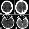
Computed Tomography with intravenous contrast: Multiple hypodense areas in the frontal and parietal lobes (black arrows - a) and in the left temporal and occipital lobe (black arrows - c). Leptomeningeal enhancement is demonstrated (grey arrow – b and d).
Fig. 3
Fig. 3
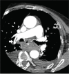
Pulmonary embolism: Computed tomography image with pulmonary embolism protocol: filling defect (white arrow) within branches of the pulmonary artery supplying the upper part of the right lower lobe.
Fig. 4
Fig. 4
Magnetic Resonance Imaging, without intravenous contrast, 7 days later: a: Fluid- attenuated Inversion Recovery (FLAIR) sequence and b: T2 sequence. Increased area of high signal intensity in the periventricular white matter, in comparison to previous MRI study.
Fig. 5
Fig. 5
Magnetic Resonance Imaging, 21 days later: a: Τ1 contrast images, b: Τ1W (weighted) and c: Susceptibility Weighted Imaging (SWI). Gyriform leptomeningeal enhancement (grey arrows-b), gyriform hyperintensities of the cortex (white arrows-a), and multiple hypointensities, cortical dot-like abnormalities (black arrows-c), indicating CNS infection with signs of recent hemorrhage.
Fig. 6
Fig. 6
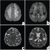
Brain MRI 21 days later a: Diffusion Weighted Imaging (DWI), b: DWI, c: Apparent Diffusion Coefficient (ADC) map (same level as -a-) and d: ADC map (same level as -b-). Decrease of restriction of diffusion in centra semiovale (white arrows – a and c) and splenium (grey arrow – b and d) in comparison to previous MRI exams.

References

    1. Kurushina OV, Barulin AE. Central Nervous System Lesions in COVID-19. Neurosci Behav Physiol. 2021;51(9):1222–1227. doi: 10.1007/s11055-021-01183-2. - DOI - PMC - PubMed
    1. Divani AA, Andalib S, Biller J, et al. Central Nervous System Manifestations Associated with COVID-19. Curr Neurol Neurosci Rep. 2020;20(12):60. doi: 10.1007/s11910-020-01079-7. Erratum in: Curr Neurol Neurosci Rep. 2020;20(12):66. - DOI - PMC - PubMed
    1. Nazari S, Azari Jafari A, Mirmoeeni S, et al. Central nervous system manifestations in COVID-19 patients: A systematic review and meta-analysis. Brain Behav. 2021;11(5):e02025. doi: 10.1002/brb3.2025. - DOI - PMC - PubMed
    1. Giorgianni A, Vinacci G, Agosti E, et al. Transient acute-onset tetraparesis in a COVID-19 patient. Spinal Cord. 2020;58(9):1042–1044. doi: 10.1038/s41393-020-0493-8. Erratum in: Spinal Cord. 2020. - DOI - PMC - PubMed
    1. Poyiadji N, Shahin G, Noujaim D, et al. COVID-19-associated Acute Hemorrhagic Necrotizing Encephalopathy: Imaging Features. Radiology. 2020;296(2):E119–E120. doi: 10.1148/radiol.2020201187. - DOI - PMC - PubMed

Publication types

LinkOut - more resources