

Mutations in the CYP27B1 gene cause vitamin D dependent rickets in pugs
- PMID: 37293695
- PMCID: PMC10365047
- DOI: 10.1111/jvim.16791
Mutations in the CYP27B1 gene cause vitamin D dependent rickets in pugs
Abstract
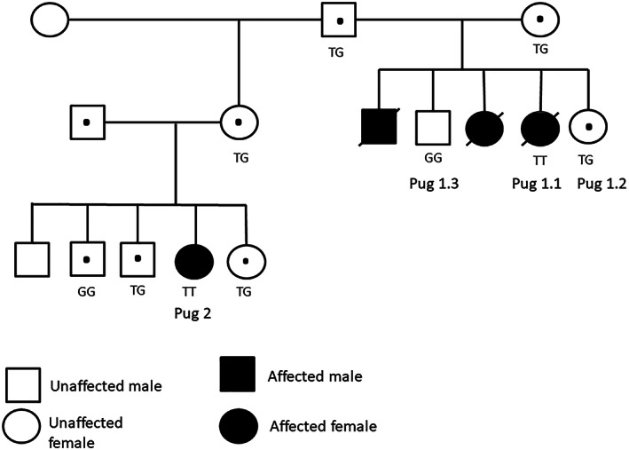
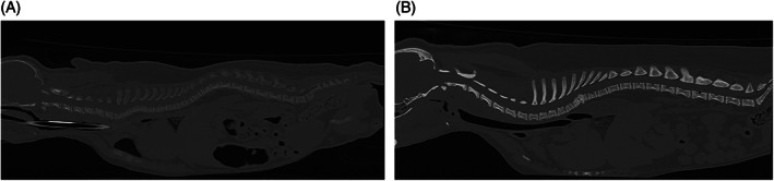
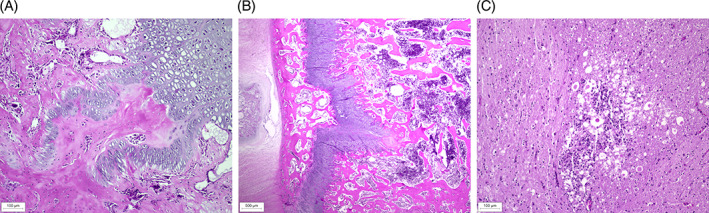
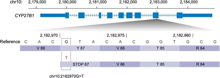
Rickets is a disorder of bone development and can be the result of either dietary or genetic causes. Here, related pugs from 2 litters were included. Three pugs had clinical signs including, lameness, bone deformities, and dyspnea. One other pug was found dead. Radiographs of 2 affected pugs, 5 and 6 months old, showed generalized widening, and irregular margination of the physes of both the appendicular and the axial skeleton with generalized decrease in bone opacity and bulbous swelling of the costochondral junctions. Two pugs had low serum calcium and 1,25 (OH)2 D3 concentrations. Test results further indicated secondary hyperparathyroidism with adequate concentrations of 25-hydroxyvitamin D. Necropsy revealed tongue-like projections of cartilage extending into the metaphysis consistent with rickets, loss of metaphyseal mineralization and lung pathology. Vitamin D-dependent rickets was diagnosed. A truncating mutation in the 1α-hydroxylase gene (CYP27B1) was identified by genome sequence analysis of the pugs with VDDR type 1A. Vitamin D-dependent rickets type 1A can occur in young pugs, and if left untreated is a life-threatening condition. Early medical intervention can reverse clinical signs and should be instituted as soon as possible.
Keywords: bone; hypocalcemia; pug; vitamin D.
© 2023 The Authors. Journal of Veterinary Internal Medicine published by Wiley Periodicals LLC on behalf of American College of Veterinary Internal Medicine.
Conflict of interest statement
Authors declare no conflict of interest.
Figures

References
-
- Bennett D. Nutrition and bone disease in the dog and cat. Vet Rec. 1976;98:313‐321. - PubMed
-
- Dittmer KE, Thompson KG. Vitamin D metabolism and rickets in domestic animals: a review. Vet Pathol. 2010;48:389‐407. - PubMed
-
- Taylor MB, Geiger DA, Saker KE, Larson MM. Diffuse osteopenia and myelopathy in a puppy fed a diet composed of an organic premix and raw ground beef. J Am Vet Med Assoc. 2009;234:1041‐1048. - PubMed
-
- Zafalon RVA, Risolia LW, Pedrinelli V, et al. Vitamin D metabolism in dogs and cats and its relation to diseases not associated with bone metabolism. J Anim Physiol Anim Nutr (Berl). 2020;104:322‐342. - PubMed
-
- Parker VJ, Rudinsky AJ, Chew DJ. Vitamin D metabolism in canine and feline medicine. J Am Vet Med Assoc. 2017;250:1259‐1269. - PubMed
Publication types
MeSH terms
Substances
Grants and funding
LinkOut - more resources
Full Text Sources
Medical
