

Exploring Antibacterial Activity and Bacterial-Mediated Allotropic Transition of Differentially Coated Selenium Nanoparticles
- PMID: 37294110
- PMCID: PMC10316328
- DOI: 10.1021/acsami.3c05100
Exploring Antibacterial Activity and Bacterial-Mediated Allotropic Transition of Differentially Coated Selenium Nanoparticles
Abstract
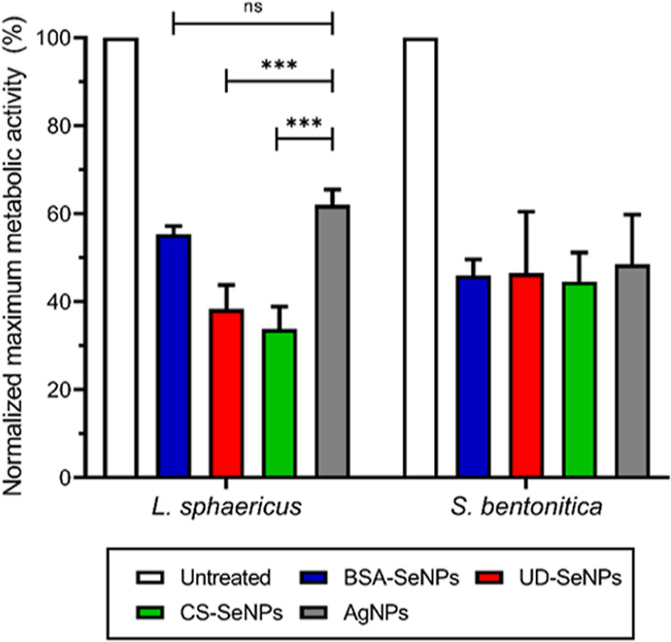
The use of metal nanoparticles (NPs) as antimicrobial agents has become a promising alternative to the problem of antibiotic-resistant bacteria and other applications. Silver nanoparticles (AgNPs) are well-known as one of the most universal biocide compounds. However, selenium nanoparticles (SeNPs) recently gained more attention as effective antimicrobial agents. This study aims to investigate the antibacterial activity of SeNPs with different surface coatings (BSA-coated, chitosan-coated, and undefined coating) on the Gram-negative Stenotrophomonas bentonitica and the Gram-positive Lysinibacillus sphaericus in comparison to AgNPs. The tested NPs had similar properties, including shape (spheres), structure (amorphous), and size (50-90 nm), but differed in their surface charge. Chitosan SeNPs exhibited a positive surface charge, while the remaining NPs assayed had a negative surface charge. We have found that cell growth and viability of both bacteria were negatively affected in the presence of the NPs, as indicated by microcalorimetry and flow cytometry. Specifically, undefined coating SeNPs displayed the highest percentage values of dead cells for both bacteria (85-91%). An increase in reactive oxygen species (ROS) production was also detected. Chitosan-coated and undefined SeNPs caused the highest amount of ROS (299.7 and 289% over untreated controls) for S. bentonitica and L. sphaericus, respectively. Based on DNA degradation levels, undefined-SeNPs were found to be the most hazardous, causing nearly 80% DNA degradation. Finally, electron microscopy revealed the ability of the cells to transform the different SeNP types (amorphous) to crystalline SeNPs (trigonal/monoclinical Se), which could have environmentally positive implications for bioremediation purposes and provide a novel green method for the formation of crystalline SeNPs. The results obtained herein demonstrate the promising potential of SeNPs for their use in medicine as antimicrobial agents, and we propose S. bentonitica and L. sphaericus as candidates for new bioremediation strategies and NP synthesis with potential applications in many fields.
Keywords: antibiotic; applications; bioremediation; nanoparticles; selenium.
Conflict of interest statement
The authors declare no competing financial interest.
Figures


Similar articles
-
Development of nanocomposite-selenium filter for water disinfection and bioremediation of wastewater from Hg and AgNPs.Sci Rep. 2024 Sep 13;14(1):21443. doi: 10.1038/s41598-024-70120-3. Sci Rep. 2024. PMID: 39271750 Free PMC article.
-
Dual-stabilized selenium nanoparticles with chitosan and SS31 peptide: Resolving instability for enhancing ROS elimination, suppressing inflammation, and combating bacterial infections.Colloids Surf B Biointerfaces. 2025 Sep;253:114749. doi: 10.1016/j.colsurfb.2025.114749. Epub 2025 Apr 30. Colloids Surf B Biointerfaces. 2025. PMID: 40318392
-
Green and ecofriendly biosynthesis of selenium nanoparticles using Urtica dioica (stinging nettle) leaf extract: Antimicrobial and anticancer activity.Biotechnol J. 2022 Feb;17(2):e2100432. doi: 10.1002/biot.202100432. Epub 2021 Nov 21. Biotechnol J. 2022. PMID: 34747563
-
Biogenesis of Selenium Nanoparticles Using Green Chemistry.Top Curr Chem (Cham). 2017 Nov 9;375(6):88. doi: 10.1007/s41061-017-0176-x. Top Curr Chem (Cham). 2017. PMID: 29124492 Review.
-
Application of Selenium Nanoparticles in Localized Drug Targeting for Cancer Therapy.Anticancer Agents Med Chem. 2022 Aug 4;22(15):2715-2725. doi: 10.2174/1871520622666220215122756. Anticancer Agents Med Chem. 2022. PMID: 35168523 Review.
Cited by
-
A Se Nanoparticle/MgFe-LDH Composite Nanosheet as a Multifunctional Platform for Osteosarcoma Eradication, Antibacterial and Bone Reconstruction.Adv Sci (Weinh). 2024 Sep;11(33):e2403791. doi: 10.1002/advs.202403791. Epub 2024 Jul 3. Adv Sci (Weinh). 2024. PMID: 38958509 Free PMC article.
-
Emerging Biohybrids of Aptamer-Based Nano-Biosensing Technologies for Effective Early Cancer Detection.Mol Diagn Ther. 2024 Jul;28(4):425-453. doi: 10.1007/s40291-024-00717-x. Epub 2024 May 22. Mol Diagn Ther. 2024. PMID: 38775897 Review.
-
Targeting Spore-Forming Bacteria: A Review on the Antimicrobial Potential of Selenium Nanoparticles.Foods. 2024 Dec 12;13(24):4026. doi: 10.3390/foods13244026. Foods. 2024. PMID: 39766969 Free PMC article. Review.
-
Nanotechnology's frontier in combatting infectious and inflammatory diseases: prevention and treatment.Signal Transduct Target Ther. 2024 Feb 21;9(1):34. doi: 10.1038/s41392-024-01745-z. Signal Transduct Target Ther. 2024. PMID: 38378653 Free PMC article. Review.
-
Bioinspired Superhydrophobic Nanocoating Based on Polydopamine and Nanodiamonds to Mitigate Bacterial Attachment to Polyvinyl Chloride Surfaces in Food Industry Environments.Ind Eng Chem Res. 2024 Mar 27;63(14):6235-6248. doi: 10.1021/acs.iecr.3c04230. eCollection 2024 Apr 10. Ind Eng Chem Res. 2024. PMID: 38617109 Free PMC article.
References
-
- Kolahalam L. A.; Viswanath I. V. K.; Diwakar B. S.; Govindh B.; Reddy V.; Murthy Y. L. N. Review on Nanomaterials: Synthesis and Applications. Mater. Today Proc. 2019, 18, 2182–2190. 10.1016/j.matpr.2019.07.371. - DOI
-
- Karami A.; Monsef R.; Shihan M. R.; Qassem L. Y.; Falah M. W.; Salavati-Niasari M. UV-light-induced Photocatalytic Response of Pechini sol–gel Synthesized Erbium Vanadate Nanostructures toward Degradation of Colored Pollutants. Environ. Technol. Innov. 2022, 28, 102947.10.1016/j.eti.2022.102947. - DOI
-
- Brandelli A.; Ritter A. C.; Veras F. F.. Antimicrobial Activities of Metal Nanoparticles. In Metal Nanoparticles In Pharma; Rai P. D. M., Shegokar P. D. R., Eds.; Springer International Publishing: Cham, 2017; pp 337–363.
MeSH terms
Substances
LinkOut - more resources
Full Text Sources
Molecular Biology Databases
Miscellaneous
