

Diurnal functional and anatomical changes in X-linked retinoschisis
- PMID: 37294434
- PMCID: PMC10587233
- DOI: 10.1007/s00417-023-06106-0
Diurnal functional and anatomical changes in X-linked retinoschisis
Abstract
Background: To investigate the changes in macular cystic schisis (MCS) and sensitivity during the day in X-linked retinoschisis (XLRS) patients.
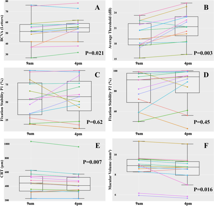
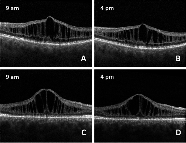
Methods: Treatment-naïve patients with genetically verified XLRS underwent best-correlated visual acuity (BCVA) testing with ETDRS charts, spectral domain optical coherence tomography, and microperimetry (MP) twice a day, at 9 a.m. and 4 p.m., to measure changes in central retinal thickness (CRT), macular volume (MV), average threshold (AT), and fixation stability parameters (P1 and P2).
Results: At baseline, the BCVA of the 14 eyes of 8 patients amounted 0.73 (± 0.23) LogMAR. Between timepoints, the BCVA increased in 3.21 letters (p = .021), the AV improved in 1.84 dB (p = .03, 9.73%), the CRT decreased in 24.43 µm (p = .007, - 4.05%), and the MV dropped in 0.27 µm3 (p = .016, - 2.68%). P1 and P2 did not variate. The collapse of the MCS led to the reduction of macula thickness. CRT at baseline correlated with the decrease of CRT (Spearman's ρ: - 0.83 [p = .001]). Age and change of BCVA, CRT, and AV did not correlate among one another. Eyes with disrupted ellipsoid zone showed a more prominent change in CRT (p = .050). Photoreceptor outer segment length and integrity of the external limiting membrane and cone outer segment tips were not associated with BCVA, AT, or CRT variation.
Conclusion: Eyes of treatment-naïve XLRS patients show diurnal macular thickness and function changes. Eyes with pronounced macular thickness show a greater reduction of the MCS. These results should be taken into consideration in upcoming clinical trials in XLRS.
Trial registration number: Institutional Review Board of the Hamburg Medical Chamber (Ethik-Kommission der Ärztekammer Hamburg): 2020-10,328.
Keywords: Diurnal variation; Microperimetry; Optical coherence tomography; RS1 gene mutation; Retinoschisis; X-linked retinoschisis.
© 2023. The Author(s).
Conflict of interest statement
The authors declare no competing interests.
Figures
References
LinkOut - more resources
Full Text Sources
Research Materials
Miscellaneous
