

Foveal Eversion is Associated with High Persistence of Macular Edema and Visual Acuity Deterioration in Retinal Vein Occlusion
- PMID: 37294523
- PMCID: PMC10287597
- DOI: 10.1007/s40123-023-00734-9
Foveal Eversion is Associated with High Persistence of Macular Edema and Visual Acuity Deterioration in Retinal Vein Occlusion
Abstract
Introduction: Foveal eversion (FE) is a recently described optical coherence tomography (OCT) finding associated with negative outcome in diabetic macular edema. The main goal of the present study was to investigate the role of the FE metric in the diagnostic workup of retinal vein occlusion (RVO).
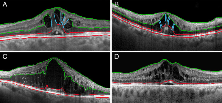
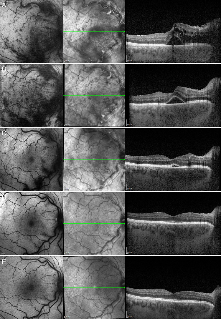
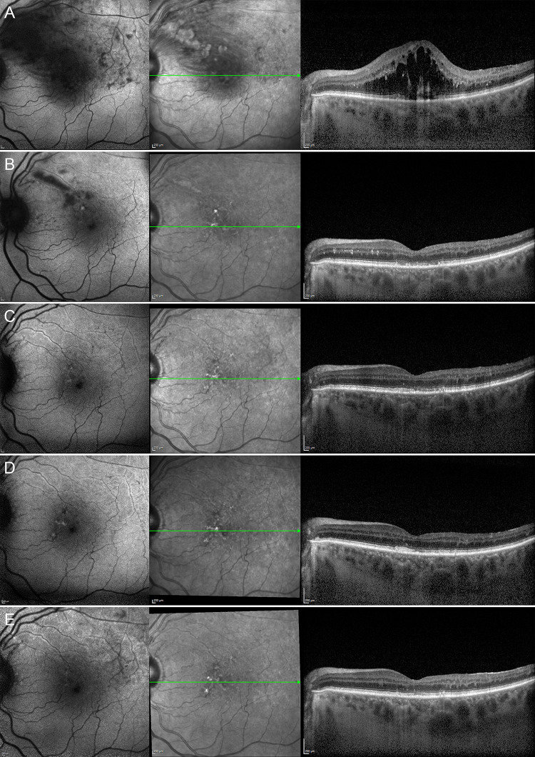
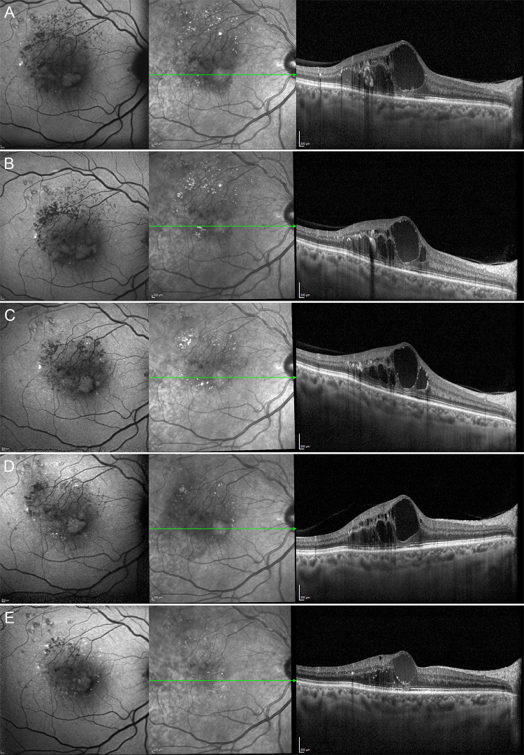
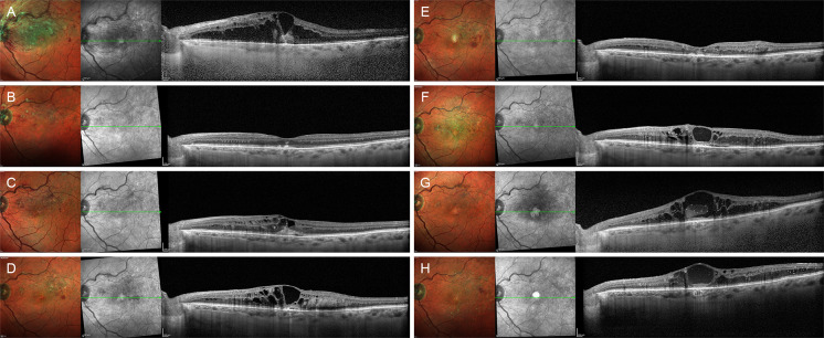
Methods: This study was a retrospective, observational case series. We included 168 eyes (168 patients) affected by central RVO (CRVO) and 116 eyes (116 patients) affected by branch (RVO). We collected clinical and imaging data from CRVO and BRVO eyes affected by macular edema with a minimum follow-up of 12 months. On structural OCT, we classified FE as pattern 1a, characterized by thick vertical intraretinal columns, pattern 1b, presenting thin vertical intraretinal lines, and pattern 2, showing no signs of vertical lines in the context of the cystoid macular edema. For statistical purposes, we considered data collected at baseline, after 1 year and at the last follow-up.
Results: The mean follow-up was 40 ± 25 months for CRVO eyes and 36 ± 24 months for BRVO eyes. We found FE in 64 of 168 CRVO eyes (38%) and in 25 of 116 BRVO eyes (22%). Most of the eyes developed FE during the follow-up. For CRVO eyes, we found 6 eyes (9%) with pattern 1a, 17 eyes (26%) with pattern 1b and 41 eyes (65%) with pattern 2. Of those BRVO eyes with FE, we found 8 eyes (32%) with pattern 1a + 1b and 17 eyes (68%) with pattern 2. In both CRVO and BRVO the presence of FE was significantly associated with higher persistence of macular edema and worse outcome, with FE pattern 2 representing the most severe condition. Remarkably, FE patterns 1a and 1b were characterized by BCVA stability over the follow-up, whereas FE pattern 2 showed significant bestcorrected visual acuity (BCVA) worsening at the end of the follow-up.
Conclusions: FE can be considered a negative prognostic biomarker in RVO, associated with higher persistence of macular edema and worse visual outcome. Müller cell impairment might represent the pathogenic mechanism leading to the loss of macular structural support and impairment of fluid homeostasis.
Keywords: BRVO; CRVO; Foveal eversion; Macular edema; OCT; Retinal vein occlusion.
© 2023. The Author(s).
Conflict of interest statement
Francesco Bandello has consulted for: Alcon (Fort Worth, TX, USA), Alimera Sciences (Alpharetta, GA, USA), Allergan Inc (Irvine, CA, USA), Farmila-Thea (Clermont-Ferrand, France), Bayer Shering-Pharma (Berlin, Germany), Bausch And Lomb (Rochester, NY, USA), Genentech (San Francisco, CA, USA), Hoffmann-La-Roche (Basel, Switzerland), NovagaliPharma (Évry, France), Novartis (Basel, Switzerland), Sanofi-Aventis (Paris, France), Thrombogenics (Heverlee, Belgium), Zeiss (Dublin, USA). Alessandro Arrigo, Emanuela Aragona, Alessio Antropoli, Lorenzo Bianco, Andrea Rosolia, Andrea Saladino and Maurizio Battaglia Parodi have nothing to disclose.
Figures
Similar articles
-
Longitudinal Assessment of the Choroidal Vascularity Index in Eyes with Branch Retinal Vein Occlusion-Associated Cystoid Macular Edema.Ophthalmol Ther. 2023 Aug;12(4):2103-2115. doi: 10.1007/s40123-023-00731-y. Epub 2023 May 23. Ophthalmol Ther. 2023. PMID: 37221425 Free PMC article.
-
Collateral Vessel Development in Central and Branch Retinal Vein Occlusions Are Associated With Worse Visual and Anatomic Outcomes.Invest Ophthalmol Vis Sci. 2021 Nov 1;62(14):1. doi: 10.1167/iovs.62.14.1. Invest Ophthalmol Vis Sci. 2021. PMID: 34724540 Free PMC article.
-
[Analysis of visual prognosis and correlative factors in retinal vein occlusion].Zhonghua Yan Ke Za Zhi. 2002 Feb;38(2):98-102. Zhonghua Yan Ke Za Zhi. 2002. PMID: 11955310 Chinese.
-
Photocoagulation for retinal vein occlusion.Prog Retin Eye Res. 2021 Nov;85:100964. doi: 10.1016/j.preteyeres.2021.100964. Epub 2021 Mar 11. Prog Retin Eye Res. 2021. PMID: 33713810 Review.
-
Comparison between anti-VEGF therapy and corticosteroid or laser therapy for macular oedema secondary to retinal vein occlusion: A meta-analysis.J Clin Pharm Ther. 2017 Oct;42(5):519-529. doi: 10.1111/jcpt.12551. Epub 2017 Jun 22. J Clin Pharm Ther. 2017. PMID: 28639290 Review.
Cited by
-
Digital histology of retinal microaneurysms as provided by dense B-scan (DART) OCTA: characteristics and clinical relevance in diabetic retinopathy.Eye (Lond). 2024 Nov;38(16):3108-3112. doi: 10.1038/s41433-024-03230-x. Epub 2024 Jul 15. Eye (Lond). 2024. PMID: 39009799
-
What is Occluding Our Understanding of Retinal Vein Occlusion?Ophthalmol Ther. 2024 Dec;13(12):3025-3034. doi: 10.1007/s40123-024-01042-6. Epub 2024 Oct 10. Ophthalmol Ther. 2024. PMID: 39387960 Free PMC article. No abstract available.
-
Proteome Analysis of Bevacizumab Intervention in Experimental Central Retinal Vein Occlusion.J Pers Med. 2023 Nov 7;13(11):1580. doi: 10.3390/jpm13111580. J Pers Med. 2023. PMID: 38003895 Free PMC article.
References
LinkOut - more resources
Full Text Sources
