. 2023 Jun 7;15(11):4625-4641.
doi: 10.18632/aging.204755. Epub 2023 Jun 7.

Short telomeres in alveolar type II cells associate with lung fibrosis in post COVID-19 patients with cancer

Affiliations

Short telomeres in alveolar type II cells associate with lung fibrosis in post COVID-19 patients with cancer

Paula Martínez et al. Aging (Albany NY). .

Abstract

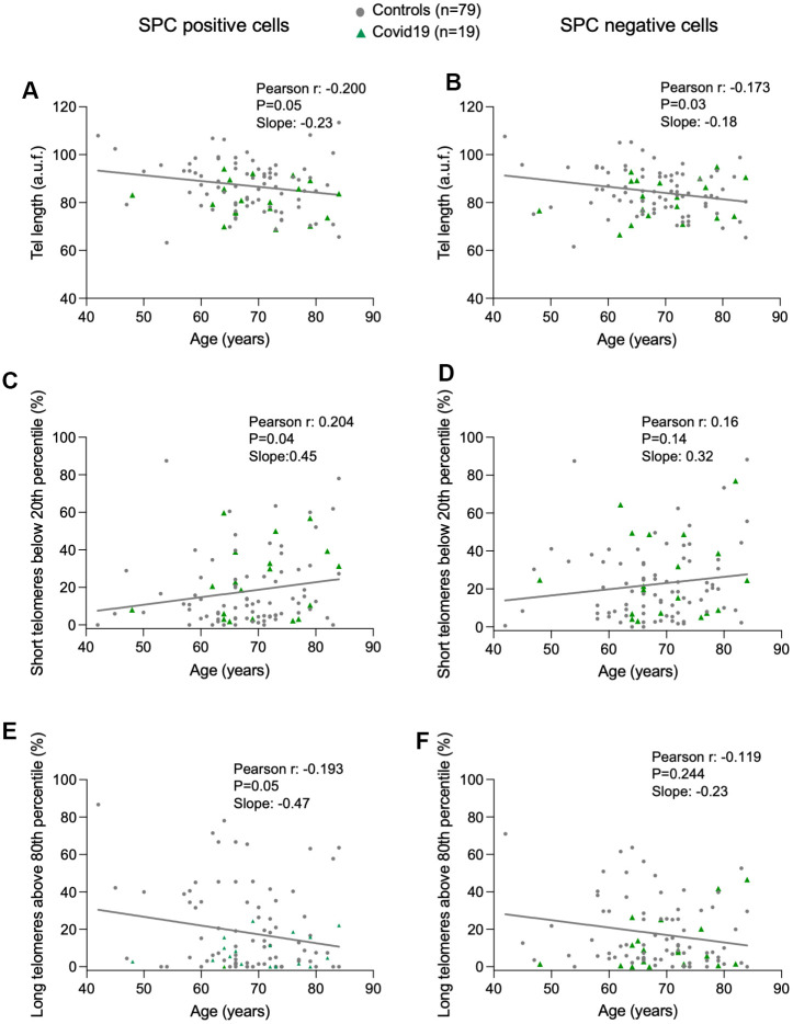
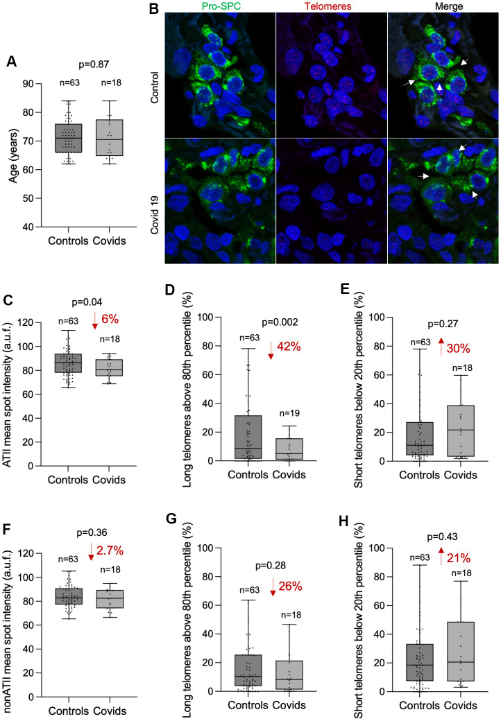
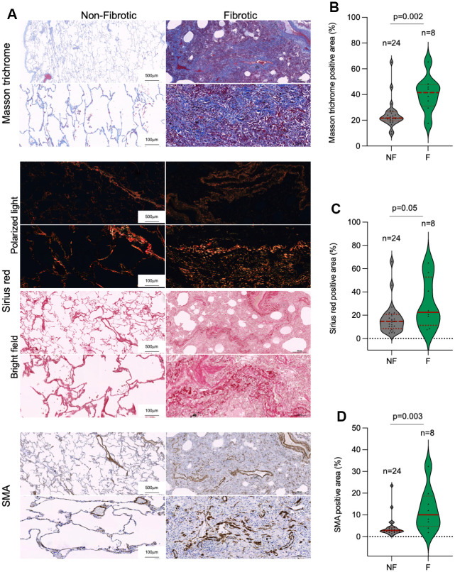
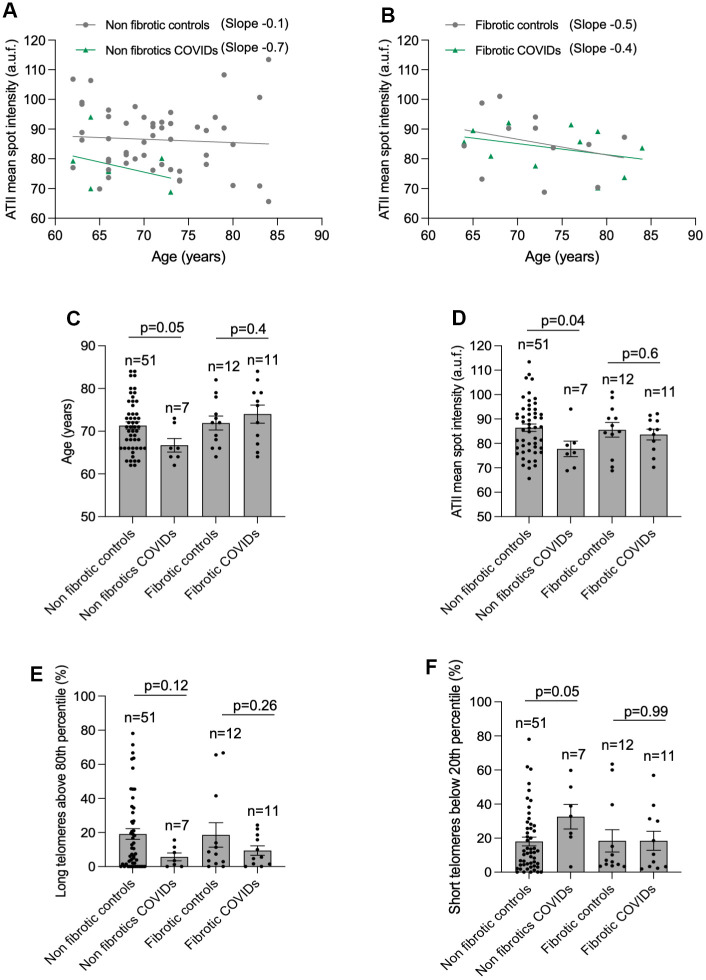
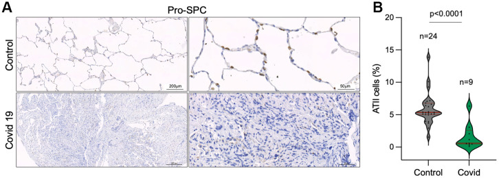
The severe acute respiratory syndrome coronavirus 2 (SARS-CoV-2) is responsible for the coronavirus disease 2019 (COVID-19) pandemic. The severity of COVID-19 increases with each decade of life, a phenomenon that suggest that organismal aging contributes to the fatality of the disease. In this regard, we and others have previously shown that COVID-19 severity correlates with shorter telomeres, a molecular determinant of aging, in patient's leukocytes. Lung injury is a predominant feature of acute SARS-CoV-2 infection that can further progress to lung fibrosis in post-COVID-19 patients. Short or dysfunctional telomeres in Alveolar type II (ATII) cells are sufficient to induce pulmonary fibrosis in mouse and humans. Here, we analyze telomere length and the histopathology of lung biopsies from a cohort of alive post-COVID-19 patients and a cohort of age-matched controls with lung cancer. We found loss of ATII cellularity and shorter telomeres in ATII cells concomitant with a marked increase in fibrotic lung parenchyma remodeling in post- COVID-19 patients compared to controls. These findings reveal a link between presence of short telomeres in ATII cells and long-term lung fibrosis sequel in Post-COVID-19 patients.

Keywords: ATII cells; COVID-19; SARS-CoV2; lung fibrosis; telomeres.


Conflict of interest statement

CONFLICTS OF INTEREST: The authors declare that they have no conflicts of interest.

Figures

Figure 1
Figure 1
Progressive telomere shortening with age in lung cells. (AF) Linear regression and Pearson correlation analyses between telomere intensity in pro-SPC positive (A) and pro-SPC negative cells (B), between percentage of short telomeres in pro-SPC positive (C) and pro-SPC negative cells (D), and between percentage of long telomeres in pro-SPC positive (E) and pro-SPC negative cells (F) and age in lung section from patients under study including control and COVID-19 samples. The Pearson r coefficient and the P value are indicated.
Figure 2
Figure 2
COVID-19 patients present shorter telomeres in the alveolar type II cells than controls. (A) Box and Whisker plot representation of control and COVID-19 patients’ age within 62 and 84 years old. Differences in age distributions between both control and COVID-19 sample groups was analyzed by Kolmogorov-Smirnov test. The p-value is indicated. (B) Representative images of telomere quantitative fluorescence in situ hybridization (q-FISH) combined with immunofluorescence against Pro-SPC in a control and a COVID-19 lung samples. (CH) Box and Whisker plot representation of mean telomeric spot intensity per nucleus (C, F), percentage of long telomeres above 80th percentile (D, G) and percentage of short telomeres below 20th percentile (E, H) in alveolar type II (ATII) (CE) and in non-ATII cells (FH) in lung sections from control and COVID-19 patients within 62 and 84 year-old age interval. Statistical significance in (CH) was assessed using unpaired Student’s t test with Welch’s correction and the p-value is indicated. The percent increase or decrease between control and COVID-19 samples are indicated in each plot.
Figure 3
Figure 3
Collagen depots in patients diagnosed with lung fibrosis post-surgery. (A) Representative images of Masson trichrome, Sirius red (polarized light and bright field) and smooth muscle actine (SMA) staining in non-fibrotic and fibrotic lungs. (BD) Quantification of Masson trichrome (B), Sirius red (C) and SMA (D) positive stained lung area in non-fibrotic (NF) and fibrotic (F) lung samples. The samples analyzed correspond to Control 1-23 and COVID-19 1-9 (Table 1). Statistical significance was assessed using Student’s t test and the p-value is indicated.
Figure 4
Figure 4
Higher rate of telomere shortening with age in COVID-19 patients. (A, B) Linear regression analyses between telomere intensity in pro-SPC positive cells and age in lung section from control and COVID-19 patients not presenting lung fibrosis (A) or being histopathologically diagnosed with lung fibrosis (B) post-surgery. The slope of the linear regression is shown. (C) Mean age of control and COVID-19 patients within 62 and 84 year-old interval presenting or not lung fibrosis post-surgery. (DF) Mean telomeric spot intensity per nucleus (D), percentage of long telomeres above 80th percentile (E) and percentage of short telomeres below 20th percentile (F) in alveolar type II in lung sections from control and COVID-19 patients within 62 and 84 year-old age interval. Statistical significance in (CF) was assessed using unpaired Student’s t test and the p-value is indicated.
Figure 5
Figure 5
Decreased number of alveolar type II cells in the lungs of COVID-19 patients. (A, B) Representative images of prosurfactant protein C (pro-SPC) immunostaining, a marker of alveolar type II cells (ATII) (A) and quantification of pro-SpC positive cells in the lungs of control and COVID-19 patients (B) Statistical significance was assessed using Student’s t test and the p-value is indicated.

Similar articles

Cited by

References

    1. Knudsen L, Ochs M. The micromechanics of lung alveoli: structure and function of surfactant and tissue components. Histochem Cell Biol. 2018; 150:661–76. 10.1007/s00418-018-1747-9 - DOI - PMC - PubMed
    1. Olajuyin AM, Zhang X, Ji HL. Alveolar type 2 progenitor cells for lung injury repair. Cell Death Discov. 2019; 5:63. 10.1038/s41420-019-0147-9 - DOI - PMC - PubMed
    1. Herzog EL, Brody AR, Colby TV, Mason R, Williams MC. Knowns and unknowns of the alveolus. Proc Am Thorac Soc. 2008; 5:778–82. 10.1513/pats.200803-028HR - DOI - PMC - PubMed
    1. de Lange T. Shelterin: the protein complex that shapes and safeguards human telomeres. Genes Dev. 2005; 19:2100–10. 10.1101/gad.1346005 - DOI - PubMed
    1. Martínez P, Blasco MA. Telomeric and extra-telomeric roles for telomerase and the telomere-binding proteins. Nat Rev Cancer. 2011; 11:161–76. 10.1038/nrc3025 - DOI - PubMed

Publication types