Structure of rosettes in the zona glomerulosa of human adrenal cortex
- PMID: 37294692
- PMCID: PMC10485581
- DOI: 10.1111/joa.13912
Structure of rosettes in the zona glomerulosa of human adrenal cortex
Abstract
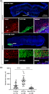
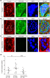
Recent studies in mouse models have demonstrated that the multi-cellular rosette structure of the adrenal zona glomerulosa (ZG) is crucial for aldosterone production by ZG cells. However, the rosette structure of human ZG has remained unclear. The human adrenal cortex undergoes remodeling during aging, and one surprising change is the occurrence of aldosterone-producing cell clusters (APCCs). It is intriguing to know whether APCCs form a rosette structure like normal ZG cells. In this study, we investigated the rosette structure of ZG in human adrenal with and without APCCs, as well as the structure of APCCs. We found that glomeruli in human adrenal are enclosed by a laminin subunit β1 (lamb1)-rich basement membrane. In slices without APCCs, each glomerulus contains an average of 11 ± 1 cells. In slices with APCCs, each glomerulus in normal ZG contains around 10 ± 1 cells, while each glomerulus in APCCs has significantly more cells (average of 22 ± 1). Similar to what was observed in mice, cells in normal ZG or in APCCs of human adrenal formed rosettes through β-catenin- and F-actin-rich adherens junctions. The cells in APCCs form larger rosettes through enhanced adherens junctions. This study provides, for the first time, a detailed characterization of the rosette structure of human adrenal ZG and shows that APCCs are not an unstructured cluster of ZG cells. This suggests that the multi-cellular rosette structure may also be necessary for aldosterone production in APCCs.
Keywords: adherens junction; adrenal zona glomerulosa; aldosterone-producing cell clusters; rosette structure.
© 2023 Anatomical Society.
Conflict of interest statement
The authors declare that they have no conflict of interest.
Figures


References
-
- Arnold, J. (1886) Ein Beitrag zur feineren Struktur und dem Chemismus der Nebennieren. Virchows Archiv für pathologische Anatomie und Physiologie und für klinische Medizin, 35, 64–107.
-
- Blankenship, J.T. , Backovic, S.T. , Sanny, J.S. , Weitz, O. & Zallen, J.A. (2006) Multicellular rosette formation links planar cell polarity to tissue morphogenesis. Developmental Cell, 11, 459–470. - PubMed
-
- Boulkroun, S. , Samson‐Couterie, B. , Dzib, J.F. , Lefebvre, H. , Louiset, E. , Amar, L. et al. (2010) Adrenal cortex remodeling and functional zona glomerulosa hyperplasia in primary aldosteronism. Hypertension, 56, 885–892. - PubMed
-
- Deane, H.W. , Shaw, J.H. & Greep, R.O. (1948) The effect of altered sodium or potassium intake on the width and cytochemistry of the zona glomerulosa of the rat's adrenal cortex. Endocrinology, 43, 133–153. - PubMed
Publication types
MeSH terms
Substances
LinkOut - more resources
Full Text Sources

