. 2023 Jul 4;62(13):2079-2092.
doi: 10.1021/acs.biochem.3c00160. Epub 2023 Jun 9.

Rapid Kinetics of Pistol Ribozyme: Insights into Limits to RNA Catalysis

Affiliations

Rapid Kinetics of Pistol Ribozyme: Insights into Limits to RNA Catalysis

Suhyun Yoon et al. Biochemistry. .

Abstract

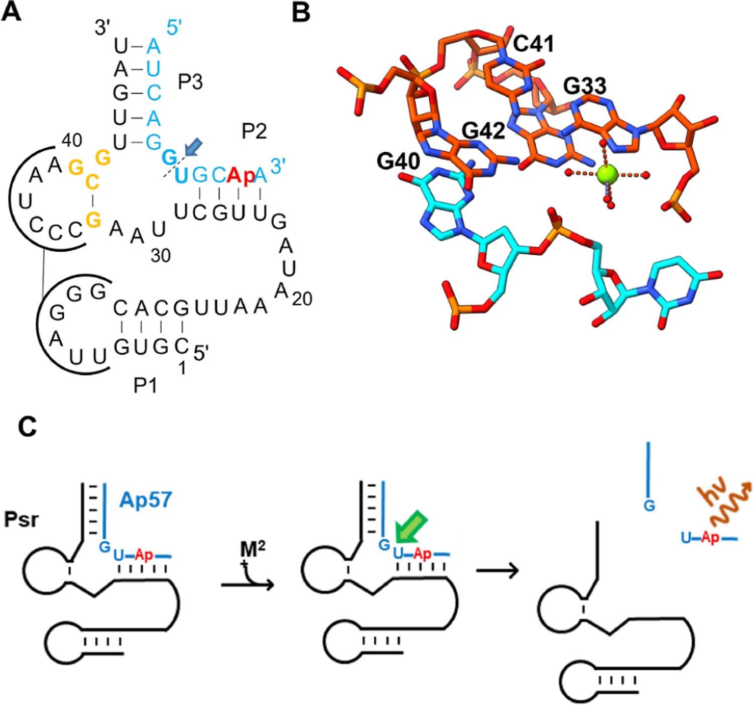
Pistol ribozyme (Psr) is a distinct class of small endonucleolytic ribozymes, which are important experimental systems for defining fundamental principles of RNA catalysis and designing valuable tools in biotechnology. High-resolution structures of Psr, extensive structure-function studies, and computation support a mechanism involving one or more catalytic guanosine nucleobases acting as a general base and divalent metal ion-bound water acting as an acid to catalyze RNA 2'-O-transphosphorylation. Yet, for a wide range of pH and metal ion concentrations, the rate of Psr catalysis is too fast to measure manually and the reaction steps that limit catalysis are not well understood. Here, we use stopped-flow fluorescence spectroscopy to evaluate Psr temperature dependence, solvent H/D isotope effects, and divalent metal ion affinity and specificity unconstrained by limitations due to fast kinetics. The results show that Psr catalysis is characterized by small apparent activation enthalpy and entropy changes and minimal transition state H/D fractionation, suggesting that one or more pre-equilibrium steps rather than chemistry is rate limiting. Quantitative analyses of divalent ion dependence confirm that metal aquo ion pKa correlates with higher rates of catalysis independent of differences in ion binding affinity. However, ambiguity regarding the rate-limiting step and similar correlation with related attributes such as ionic radius and hydration free energy complicate a definitive mechanistic interpretation. These new data provide a framework for further interrogation of Psr transition state stabilization and show how thermal instability, metal ion insolubility at optimal pH, and pre-equilibrium steps such as ion binding and folding limit the catalytic power of Psr suggesting potential strategies for further optimization.


Conflict of interest statement

The authors declare no competing financial interest.

Figures

Figure 1.
Figure 1.
Pistol ribozyme (Psr) secondary structure, active site, and overall reaction scheme. (A) Secondary structure of env25 pistol ribozyme and substrate (A57Ap) used in this study and in previous structural and mechanistic analyses. Secondary structure adapted from reference. The cleavage site is indicated by a green arrow, and the position of the 2-aminopurine modification (Ap) is shown in red. Key active site residues G40–G42 and G33 are colored orange. (B) Three-dimensional structure of the active site of Psr based on the structure of a substrate analogue containing a deoxynucleotide substitution at the cleavage site (PDB:6UEY) with the proposed interactions with active site Mg2+ ion based on a vanadate transition state mimic is highlighted. The nucleotide flanking the scissile phosphate is in blue, and key active site nucleotides are shown in orange and numbered according to the secondary structure in panel A. (C) Reaction scheme for intermolecular cleavage of the short oligonucleotide substrate (A57Ap, blue) by env25 (black). The position of the fluorescent Ap residue is red, the cleavage site is indicated by an arrow, and the increase in Ap fluorescence upon cleavage and dissociation is depicted by a star.
Figure 2.
Figure 2.
Steady-state fluorescence of Psr S and ES complexes. (A) Emission spectra of 1 μM A57Ap substrate oligonucleotide in the presence of increasing concentrations of env25 Psr (E) (λex = 305 nm). (B) Dependence of fluorescence intensity at λmax = 360 nm on the E/S ratio. Individual data points are shown as circles; the line shows a fit to the quadratic form of the equilibrium binding equation. (C) Emission spectra of S (solid orange line), E/S (dotted black line), and the E/S complex after addition of Mg2+ (blue dashed line).
Figure 3.
Figure 3.
Stopped-flow fluorescence analysis of Psr reaction kinetics. (A) Time-dependent change in A57Ap fluorescence upon mixing Psr/A57Ap and Mg2+ to achieve a final concentration of 1 μM A57Ap, 1.5 μM Psr, and 1 mM Mg2+ (open circles) or mixing with buffer without Mg2+ (filled circles). The solutions mixed in the flow cell were excited using a 300 nm LED source and total fluorescence detected using a 320 nm high pass filter. (B) Time-dependent Ap fluorescent signal after mixing of 1 μM (final concentration) E/dA57Ap complex, in which the cleavage site contains a 2′-deoxy modification, with Mg2+ to achieve a final concentration of 1 mM.
Figure 4.
Figure 4.
Temperature dependence of Psr folding and reaction kinetics. (A) CD spectra of a sample containing 1 μM dS and 1.1 μM E in 30 mM HEPES pH 8.0, 2 M NaCl, and 5 mM MgCl2 (15 °C). (B) Spectra were acquired at a series of increasing temperatures and overlaid, the change in CD signal associated with thermal unfolding is indicated by an arrow. (C) Plot of CD signal intensity at 265 nm as a function of temperature (5–85 °C). The data were fit to a two-state reversible unfolding mechanism to estimate the Tm for the Psr ES (open circles) and ES complex in 5 mM Mg2+ (filled circles). (D) Thermodynamic analysis of Psr temperature dependence (7–25 °C). The reaction rate decreases dramatically above 25 °C as evidenced by the data point obtained at 28 °C (filled circle), which was excluded from data fitting. The temperature-dependent data (open circles) are fit to a linear equation to calculate the apparent activation energy (ΔHapp = 1.36 ± 0.06 kcal/mol) and entropy change (ΔSapp = −0.66 e.u.).
Figure 5.
Figure 5.
Analysis of solvent kinetic isotope effects on Psr reaction kinetics. (A) Time-dependent change in 2Ap fluorescence upon mixing ES and Mg2+ to achieve a final concentration of 1 μM S, 1.5 μM E at pL (L = H or D) 9.4, 8.4, and 7.4 run in H2O (open symbols) or D2O (filled symbols). (B) pL profiles of log(kobs) for Psr in H2O (open symbols) and D2O (filled symbols) are shown. The solid and dashed lines represent fits to the H2O and D2O data, respectively, to a model for an enzyme with active site acid and base with pKa values pKa,A and pKa,B, respectively (eq 7).
Figure 6.
Figure 6.
Mg2+ concentration dependence of Psr cleavage rate constant under conditions of 30 mM HEPES pH 8.0, 2 M NaCl and increasing concentrations of divalent metal ion. (A) Time-dependent changes in fluorescence of Psr ES complex mixed with MgCl2 containing buffer to achieve a final concentration of 0.2 mM (red points), 2 mM (blue points), or 20 mM (green points). (B) Dependence of kobs (circles) on Mg2+ concentration. The data are fit to an equation for equilibrium binding of two independent, non-interacting metal ion binding sites that include an activating ion MA and an ion that inhibits catalysis MB as depicted in the mechanism shown in Scheme 2.
Figure 7.
Figure 7.
Comparison of the divalent metal ion dependence of Psr. (A–F) Plot of kobs determined by stopped-flow fluorescence versus concentration for (A) Mg2+, (B) Ca2+, (C) Cd2+, (D) Mn2+, (E) Co2+, and (F) Zn2+. Data were collected at pH 6.0 for all ions except Mg2+ and Ca2+ (data collected at pH 8.0). Titration data for Mg2+, Cd2+, Co2+, and Zn2+ are fit to a mechanism for binding of an activating (MA) and inactivating (MB) metal ion described in Scheme 2 (solid line). The data for reactions with Ca2+ are fit to a model for a single activating metal ion (dashed line) or anti-cooperative binding of two divalent ions (solid line). The data for reactions in which Mn2+ was the divalent ion are fit to a single site equilibrium binding model. The fitting results and errors are reported in Table 1.
Figure 8.
Figure 8.
Analysis of kc, the rate constant for catalysis the Psr metal complex (ES*M2+). (A) Comparison of the affinity of divalent ions for nucleotide monophosphate (Kd,NMP) to the observed binding affinity of MA (KMA). The value of KMA was determined by fitting the metal titration data, as shown in Figure 7. The lack of correlation reflects the apparent specificity of the Psr active site for different ions. The data for each ion are labeled. (B) Comparison of the kc values determined by fitting the full metal ion titration datasets to Scheme 2 or estimated by fitting the data at limiting ion concentration according to Scheme 3. (C) Correlation between the pKa of the metal aquo ion for each divalent metal ion versus the observed kc/KM (green, slope = 1.3), kc estimated from fitting to Scheme 2 (red, slope = 0.71), and kc estimated using kc from Scheme 3 (blue, slope = 0.96). (D−F) Comparison of the potential correlations between log(kc) and hydration free energy (ΔGhyd), radius (rion), and absolute hardness (η) of divalent metal ions used. The data for log(kc) versus hydration free energy and radius are fit to a linear function.
Figure 9.
Figure 9.
Factors that limit the ability of Psr to catalyze RNA 2′-O-transphosphorylation. A simple mechanistic model of the reaction of the Psr ES complex shows specific steps required to reach the transition state. The ES ground state ensemble is largely folded into the native structure and undergoes divalent metal ion binding (which may include inhibitory or anti-cooperative interactions), local conformational changes, active site ionization, and formation of interactions that provide αδ catalytic modes. Individual reaction steps can be subject to factors that limit their contribution to RNA strand cleavage, or which are amenable to further optimization as discussed in the text.
Scheme 1
Scheme 1
Scheme 2
Scheme 2
Scheme 3
Scheme 3

Similar articles

Cited by

References

    1. Peng H; Latifi B; Müller S; Lupták A; Chen IA Self-cleaving ribozymes: substrate specificity and synthetic biology applications. RSC Chem. Biol 2021, 2, 1370–1383. - PMC - PubMed
    1. Jimenez RM; Polanco JA; Lupták A Chemistry and Biology of Self-Cleaving Ribozymes. Trends Biochem. Sci 2015, 40, 648–661. - PMC - PubMed
    1. Micura R; Höbartner C Fundamental studies of functional nucleic acids: aptamers, riboswitches, ribozymes and DNAzymes. Chem. Soc. Rev 2020, 49, 7331–7353. - PubMed
    1. Balke D; Hieronymus R; Müller S Challenges and Perspectives in Nucleic Acid Enzyme Engineering. Adv. Biochem. Eng. Biotechnol 2020, 170, 21–35. - PubMed
    1. Ren A; Micura R; Patel DJ Structure-based mechanistic insights into catalysis by small self-cleaving ribozymes. Curr. Opin. Chem. Biol 2017, 41, 71–83. - PMC - PubMed

Publication types