. 2023 Jun 8;186(12):2672-2689.e25.
doi: 10.1016/j.cell.2023.05.019. Epub 2023 Jun 8.

Vaccine elicitation and structural basis for antibody protection against alphaviruses

Affiliations

Vaccine elicitation and structural basis for antibody protection against alphaviruses

Matthew S Sutton et al. Cell. .

Abstract

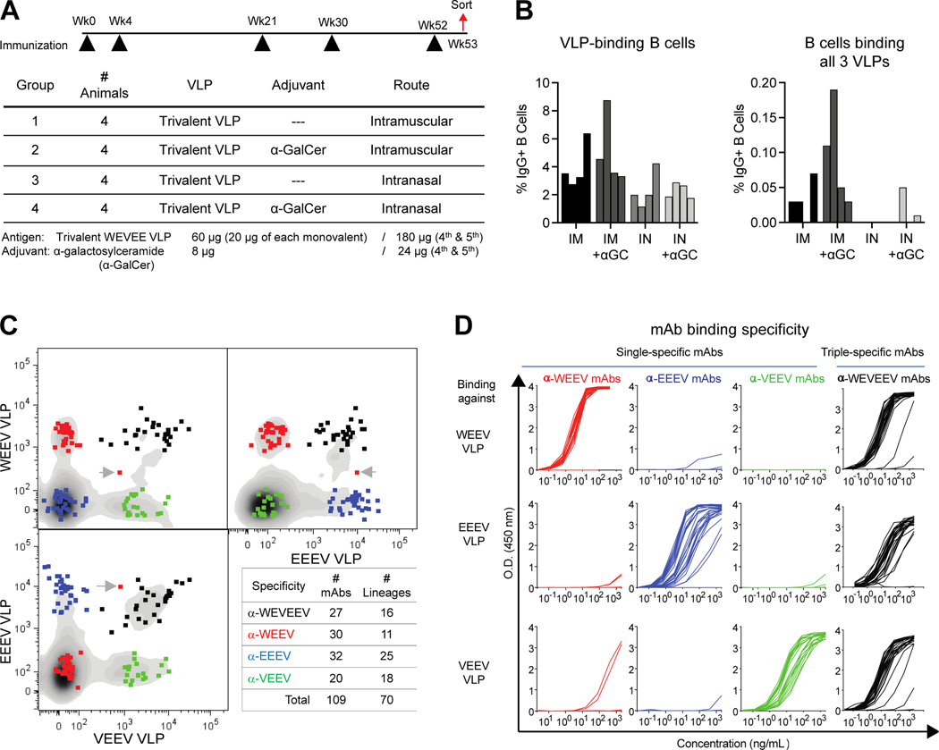
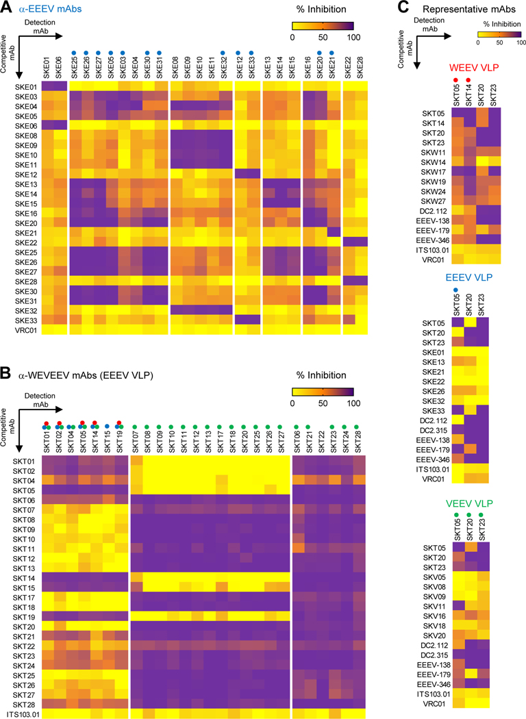
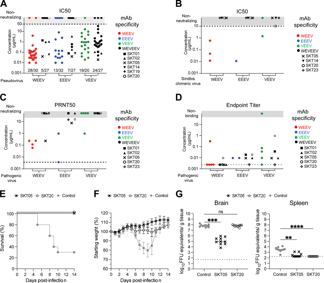
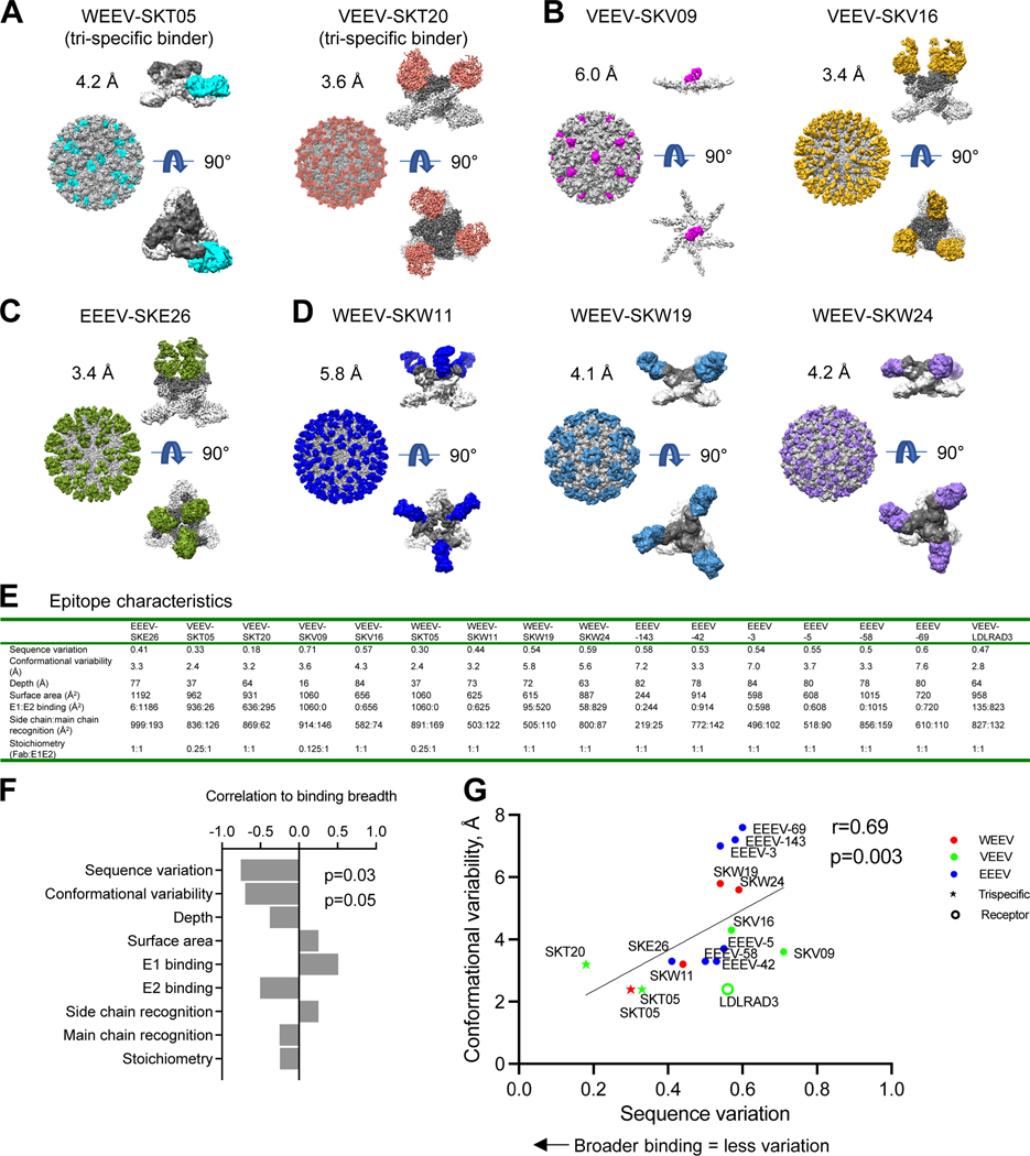
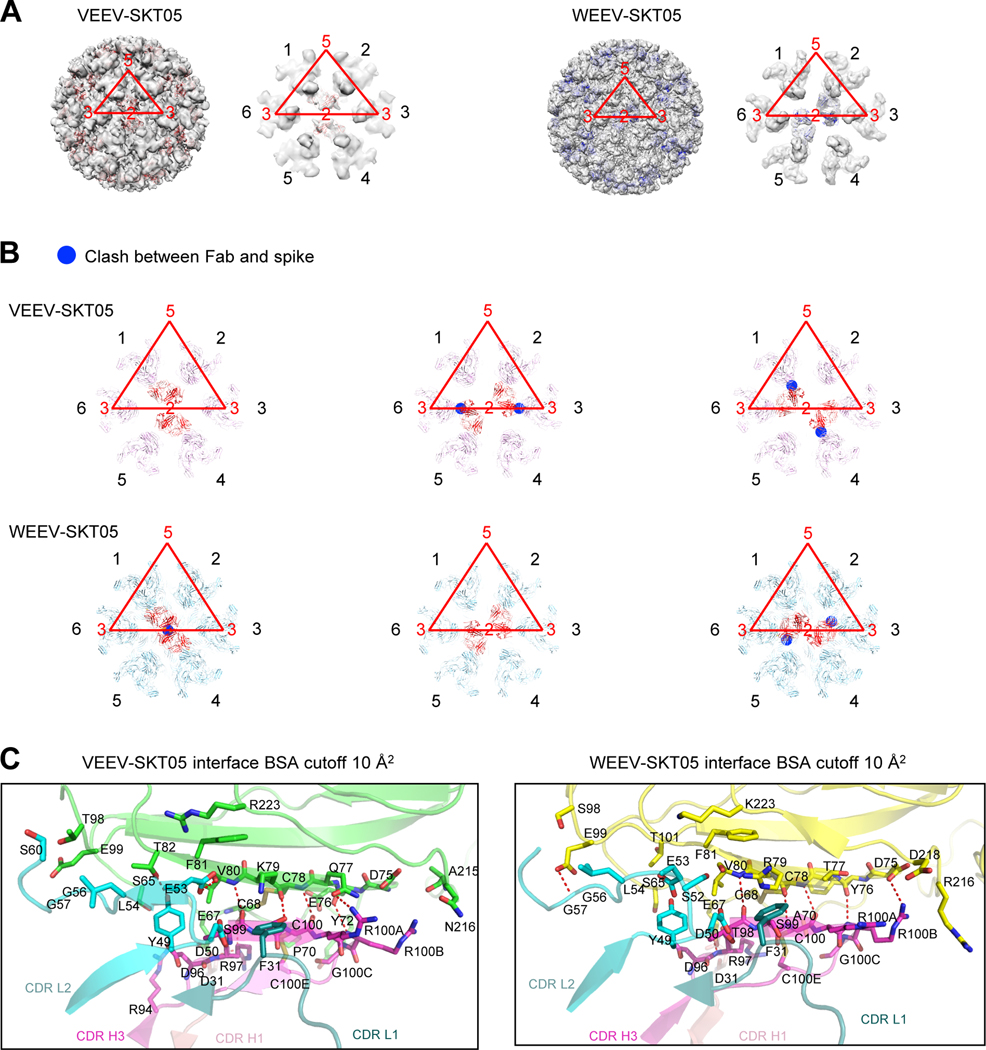
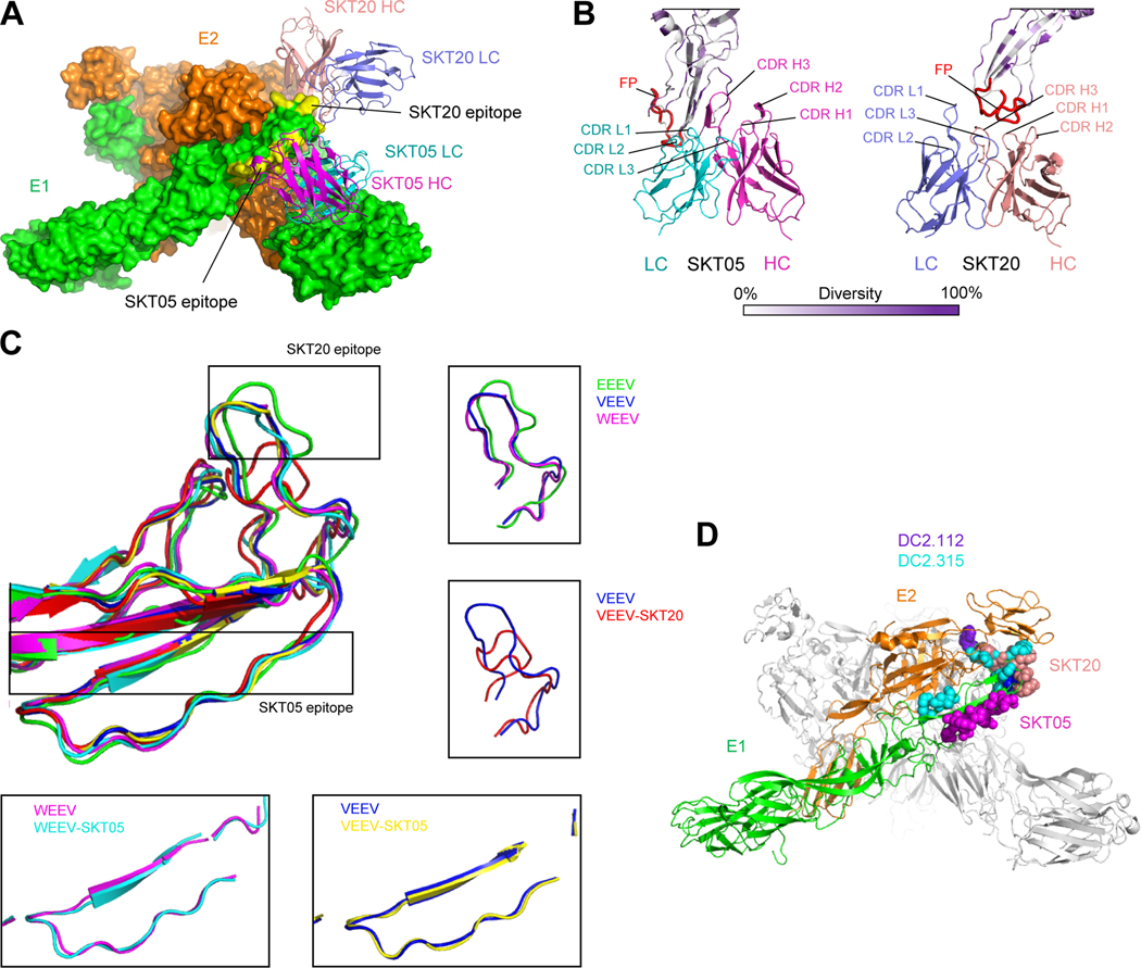
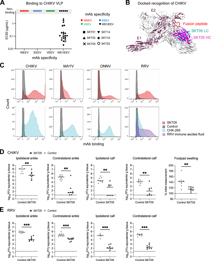
Alphaviruses are RNA viruses that represent emerging public health threats. To identify protective antibodies, we immunized macaques with a mixture of western, eastern, and Venezuelan equine encephalitis virus-like particles (VLPs), a regimen that protects against aerosol challenge with all three viruses. Single- and triple-virus-specific antibodies were isolated, and we identified 21 unique binding groups. Cryo-EM structures revealed that broad VLP binding inversely correlated with sequence and conformational variability. One triple-specific antibody, SKT05, bound proximal to the fusion peptide and neutralized all three Env-pseudotyped encephalitic alphaviruses by using different symmetry elements for recognition across VLPs. Neutralization in other assays (e.g., chimeric Sindbis virus) yielded variable results. SKT05 bound backbone atoms of sequence-diverse residues, enabling broad recognition despite sequence variability; accordingly, SKT05 protected mice against Venezuelan equine encephalitis virus, chikungunya virus, and Ross River virus challenges. Thus, a single vaccine-elicited antibody can protect in vivo against a broad range of alphaviruses.

Keywords: alphavirus; broadly neutralizing antibody; cryo-EM; in vivo challenge; vaccine.


Conflict of interest statement

Declaration of interests NIH has submitted a provisional patent application for select antibodies described in this manuscript on which M.S.S., S.K., R.V., P.D.K., and M.R. are co-inventors.

Figures

Figure 1.
Figure 1.. Trivalent VLP immunization elicits single-specific and triple-specific α-EEV mAbs in NHP.
(A) Cynomolgus macaque immunization regimen. (B) Percent of CD19+IgG+ B cells binding at least one VLP probe (left), and those binding all three VLP probes simultaneously (right). (C) Single B cells positive for binding only a single VLP (WEEV: red; EEEV: blue; VEEV: green) or all three VLPs (WEVEEV: black) were sorted into wells. A gray arrow denotes a doublet sort, comprising two B cells in one well (only one sequence was generated). Lineages were defined as having a unique V-gene allele, CDR3 sequence, and CDR3 length. (D) ELISA binding curves of single-specific and triple-specific ⍺-EEV mAbs to WEEV VLPs (top), EEEV VLPs (middle), and VEEV VLPs (bottom). Data are representative of two to three independent experiments. See also Figure S1 and Tables S1–S2.
Figure 2.
Figure 2.. Competition ELISAs identify distinct binding groups of single-specific and triple-specific α-EEV mAbs.
Competitive binding ELISAs were performed to identify overlap in EEEV VLP surface binding areas within (A) ⍺-EEEV mAbs and (B) ⍺-WEVEEV mAbs. Data were hierarchically clustered to determine competition groups for each VLP and are representative of at least two independent experiments (C) Representative single-specific mAbs, as well as previously published broadly reactive ⍺-EEV mAbs (DC2.112, DC2.315, EEEV-138, EEEV-179, and EEEV-346),, were competed against representative triple-specific mAbs. Heat maps display percent inhibition ranging from yellow (minimal competition) to orange (moderate competition) to purple (maximal competition). Negative control mAbs include either the human ⍺-HIV mAb VRC0160 or the NHP ⍺-SIV mAb ITS103.01. Colored circles above mAbs along x-axis represent the EEV pseudovirus that was neutralized (red: WEEV; blue: EEEV; green: VEEV). Data are representative of two independent experiments. See also Figures S2–S3, and Table S2.
Figure 3.
Figure 3.. Vaccine-elicited ⍺-EEV mAbs bind, neutralize, and protect against encephalitic alphavirus challenge in vivo.
(A) Neutralization IC50 values for single- and triple-specific ⍺-EEV mAbs against WEEV, EEEV, and VEEV Env-pseudotyped lentiviral reporter viruses. Fractions above x-axis indicate number of neutralizing mAbs out of total tested. Data are representative of at least two independent experiments performed in triplicate. (B) IC50 values for select ⍺-EEV mAbs against SINV-chimeric viruses. Data are representative of two independent experiments. (C) PRNT50 values for select ⍺-EEV mAbs against pathogenic WEEV (Fleming strain), EEEV (FL93–939 strain), and VEEV (TrD strain). Data are shown from one experiment after determining the appropriate starting dilution. (D) ELISA endpoint binding titers for select single-specific and triple-specific ⍺-EEV mAbs against WEEV (CBA87 strain), EEEV (FL93–939 strain), and VEEV (TrD strain). Data are shown from one experiment after determining the appropriate starting dilution. In the event an endpoint titer was not identified, results are reported as half of the lowest binding titer tested and are indicated along the dotted line. (E-G) VEEV (TC-83) challenge outcome in mice (n=10/group) that received SKT05, SKT20, or a NHP ⍺-SIV mAb as a control one day prior to inoculation. Data are representative of two independent experiments. (E) Survival rate analysis and (F) change in relative weight in mice for 14 days after inoculation. (G) Viral load was determined in the brain and spleen 5 days after inoculation. Statistical significance related to viral RNA was determined by Kruskal-Wallis test (**p< 0.0021, ***p< 0.0002, ****p< 0.0001). The dotted line indicates the limit of detection for viral RNA analysis. Colors represent mAb specificity while select triple-specific mAbs are shown as unique black symbols. Only select triple-specific mAbs were assessed in B-D, all of which had unique black symbols. See also Figure S2 and Table S2.
Figure 4.
Figure 4.. Cryo-EM structures of neutralizing antibodies with VLPs reveal that sequence variation and conformation variability inversely correlate with broad recognition.
(A-D) Cryo-EM structures of select (A) triple-specific antibodies, (B) α-VEEV antibodies, (C) α-EEEV antibodies, or (D) α-WEEV antibodies. Each antibody neutralized at least one pseudovirus. For each antibody-VLP complex, the entire complex is shown with VLP, with VLP in gray and Fabs colored. The E1E2 spike with bound Fab is also shown E1 in light gray, E2 in dark gray, and Fab colored; a side view is shown at top, and under it, a 90° rotation viewing down the spike molecular 3-fold axis (note that for VEEV-SKV09, this Fab binds directly to E1 at the 2-fold axis of the VLP, and this interaction is shown from side and along 2-fold axis). (E) Epitope characteristics are calculated for new antibodies and EEEV-143 (PDB 6xob), EEEV-42 (PDB 6mui), EEEV-3 (PDB 6mw9), EEEV-69 (PDB 6mwx), EEEV-58 (PDB 6mwv), EEEV-5 (PDB 6mwc), and LDLRAD3 (PDB 7ffn). (F) Bar graph displaying correlation of binding breadth and epitope properties. Sequence variation was calculated as a BSA-weighted average of normalized entropy. Conformational variability was calculated as a BSA-weighted average RMSD. (G) Epitope sequence and conformational variability plotted for alphavirus antibodies and receptor. Color identifies VLP with which the antibody complex structure was solved. See also Figures S4–S5 and Data S1.
Figure 5.
Figure 5.. SKT05 utilizes different symmetry to bind VEEV and WEEV.
(A) Overall view of the reconstruction density for VEEV and WEEV VLPs with bound SKT05 Fab and close-up view of icosahedral 2-fold axis surrounded with six E1:E2 spikes (labeled 1–6 in black font, with VLP symmetry axes labeled in red font, and the icosahedral asymmetric unit also shown in red). SKT05 Fabs are shown as red and blue ribbons, respectively. SKT05 bound to spikes labeled 1 and 4 in VEEV and to spikes labeled 2 and 5 in WEEV. (B) VEEV-SKT05 and WEEV-SKT05 complexes docked into spike reconstruction density surrounding 2-fold axis. Clashes are highlighted with blue circles and were observed between SKT05 and nearest polypeptide chains when VEEV-SKT05 was docked to spike pairs 2,5 and 6,3, and when WEEV-SKT05 was docked to spike pairs 1,4 and 6,3. (C) Details of interactions between VEEV and WEEV VLPs with SKT05 are shown in ribbons, with all interactions with a BSA larger than 10 Å2 shown in sticks. Left panel: interactions of VEEV E1 (green) with CDR-H1, CDR-H3, CDR-L1 and CDR-L2 of SKT05. Right panel: interactions of WEEV E1 (yellow) with CDR-H1, CDR-H3, CDR-L1 and CDR-L2 of SKT05. See also Table S3 and Data S1.
Figure 6.
Figure 6.. Structural details of SKT05 and SKT20 broad recognition.
(A) Overall view of the WEEV spike bound by SKT05 and SKT20. Molecular surfaces are shown for the E1:E2 trimer (colored green and orange, respectively), with SKT05 and SKT20 variable domains displayed in ribbons and their epitopes highlighted in yellow. (B) Close-up view of SKT20 and SKT05 in contact with WEEV E1 glycoprotein drawn in ribbons and colored by sequence diversity according to the white-to-purple key, with two conformations of the fusion peptide highlighted in red. (C) Superposition of E1 glycoproteins for EEEV, VEEV, WEEV, and their SKT05 and SKT20-bound complexes. In free EEEV, VEEV, and WEEV VLPs, E1 glycoproteins have a similar conformation for the fusion peptides. Binding of SKT05 does not affect the conformation of E1, whereas binding of SKT20 results in a dramatic change in the conformation of the fusion peptide. (D) View of the WEEV spike, displayed in ribbons representation, with one E1:E2 protomer colored green and orange, respectively, and the two other protomers colored medium and light gray. Epitopes for SKT05, SKT20, DC2.112 and DC2.315 are shown in sphere representation, and colored magenta, orange, purple, and cyan, respectively. See also Data S1.
Figure 7.
Figure 7.. SKT05 broadly recognizes and protects in vivo against arthritogenic alphaviruses.
(A) Representative single-specific mAbs, and all triple-specific mAbs, were tested for binding to chikungunya VLP. Symbol colors represent mAb specificity to the WEEV VLP (red), EEEV VLP (blue), VEEV VLP (green), or all three VLPs (black). Select triple-specific antibodies are shown as unique symbols due to their binding and neutralization profiles. Data are representative of at least two independent experiments. (B) Dock model of SKT05 and CHIKV VLP. (C) Binding to live cells infected with CHIKV, MAYV, ONNV, or RRV by SKT05 (red), CHK-265 (blue), RRV immune ascites fluid (purple), or a control mAb (gray) was determined by flow cytometry. Data are representative of three independent experiments performed in duplicate. (D-E) Arthritogenic alphavirus challenge outcome in mice (n=8/group) that received SKT05 (black) or the NHP α-SIV mAb ITS103.01 as a control (gray) one day prior to inoculation. Data are representative of two independent experiments. Viral load was determined in indicated tissues 3 days following inoculation with (D) CHIKV or (E) RRV. Footpad swelling (width x height) in the ipsilateral foot was measured prior to and 3 days following CHIKV inoculation. Statistical significance related to viral RNA was determined by a Mann-Whitney test and by unpaired t-test for footpad swelling (*p < 0.05, **p < 0.01, ***p < 0.001). The dotted line indicates the limit of detection for viral RNA analysis and the baseline foot measurement for foot swelling. See also Figures S6–S7.

Comment in

References

    1. Fox JM, and Diamond MS (2016). Immune-Mediated Protection and Pathogenesis of Chikungunya Virus. J Immunol 197, 4210–4218. 10.4049/jimmunol.1601426. - DOI - PMC - PubMed
    1. Zaid A., Burt FJ., Liu X., Poo YS., Zandi K., Suhrbier A., Weaver SC., Texeira MM., and Mahalingam S. (2021). Arthritogenic alphaviruses: epidemiological and clinical perspective on emerging arboviruses. Lancet Infect Dis 21, e123–e133. 10.1016/S1473-3099(20)30491-6. - DOI - PubMed
    1. Silva LA, and Dermody TS (2017). Chikungunya virus: epidemiology, replication, disease mechanisms, and prospective intervention strategies. J Clin Invest 127, 737–749. 10.1172/JCI84417. - DOI - PMC - PubMed
    1. Ronca SE, Dineley KT, and Paessler S. (2016). Neurological Sequelae Resulting from Encephalitic Alphavirus Infection. Front Microbiol 7, 959. 10.3389/fmicb.2016.00959. - DOI - PMC - PubMed
    1. Arechiga-Ceballos N, and Aguilar-Setien A. (2015). Alphaviral equine encephalomyelitis (Eastern, Western and Venezuelan). Rev Sci Tech 34, 491–501. 10.20506/rst.34.2.2374. - DOI - PubMed

Publication types