. 2023 Jun 15;83(12):2020-2034.e6.
doi: 10.1016/j.molcel.2023.05.025. Epub 2023 Jun 8.

Poly(A)-binding protein is an ataxin-2 chaperone that regulates biomolecular condensates

Affiliations

Poly(A)-binding protein is an ataxin-2 chaperone that regulates biomolecular condensates

Steven Boeynaems et al. Mol Cell. .

Abstract

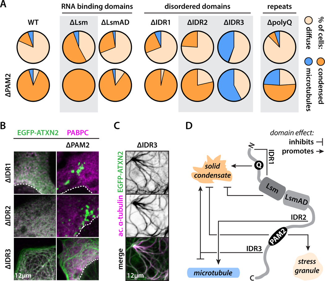
Biomolecular condensation underlies the biogenesis of an expanding array of membraneless assemblies, including stress granules (SGs), which form under a variety of cellular stresses. Advances have been made in understanding the molecular grammar of a few scaffold proteins that make up these phases, but how the partitioning of hundreds of SG proteins is regulated remains largely unresolved. While investigating the rules that govern the condensation of ataxin-2, an SG protein implicated in neurodegenerative disease, we unexpectedly identified a short 14 aa sequence that acts as a condensation switch and is conserved across the eukaryote lineage. We identify poly(A)-binding proteins as unconventional RNA-dependent chaperones that control this regulatory switch. Our results uncover a hierarchy of cis and trans interactions that fine-tune ataxin-2 condensation and reveal an unexpected molecular function for ancient poly(A)-binding proteins as regulators of biomolecular condensate proteins. These findings may inspire approaches to therapeutically target aberrant phases in disease.

Keywords: ATXN2; PABPC; amyotrophic lateral sclerosis; microtubule-binding protein; polyQ; protein aggregation; protein phase separation; short linear motif; spinocerebellar ataxia; stress granules.


Conflict of interest statement

Declaration of interests A.D.G. is a scientific founder of Maze Therapeutics. A.S.H. is a scientific consultant for Dewpoint Therapeutics and on the Scientific Advisory Board for Prose Foods.

Figures

Figure 1:
Figure 1:. The PAM2 switch regulates ATXN2’s behavior in stress and non-stress conditions.
(A) Stress granules form upon cellular stress. (B) ATXN2 is dispensable for arsenite-induced stress granule assembly. Average percentage of cells with stress granules is shown. n = 3 experiments with a total of [255–285] cells. Dashed lines highlight nuclei. EGFP is shown in inverse gray scale (see also Fig. S1A). (C) Phase separation of scaffold proteins and RNA drives stress granule assembly, with subsequent recruitment of non-essential client proteins, such as ATXN2. (D) Domain structure and disorder prediction of ATXN2. Metapredict score: 1 = disordered, 0 = folded. (E) Deletion of PAM2 prevents homogeneous partitioning of ATXN2 in stress granules. (F) Quantification of the heterogeneity of ATXN2 deletion mutant distribution within the stress granule compartment (see STAR methods). One-way ANOVA. n = 30 stress granules. (G) PAM2 deletion drives spontaneous condensation of ATXN2 into small granules under non-stress conditions (see also Fig. S1B). (H) Wildtype ATXN2 can spontaneously condense upon overexpression. ΔPAM2 ATXN2 condensation is not an overexpression artefact (see also Fig. S2). Scatterplots show cells with diffuse or condensed ATXN2 localization. Average cytoplasmic EGFP intensity. Cells combined from 3 experiments. Mann-Whitney. * p-value < 0.05, **** p-value < 0.0001. Panel (B) shows HeLa cells. All other panels show U2OS cells. Every picture shows endogenous PABPC staining. EGFP-ATXN2 (mutants) are expressed from a plasmid.
Figure 2:
Figure 2:. The PAM2 switch is functionally conserved across eukaryotes.
(A) Eukaryote tree highlighting different clades and model organisms. Archaea are the outgroup. Black stars indicate organisms compared in this figure. (B) PAM2 deletion results in the formation of a single large CoATXN2 granule upon expression in C. owczarzaki, opposed to multiple small wildtype granules. N denotes nucleus (see also Fig. S3B–D). (C) TbATXN2 localizes diffusely in trypanosomes, whereas the ΔPAM2 mutant spontaneously condenses (see also Fig. S3E). N denotes nucleus, K denotes kinetoplast. (D) AtATXN2 localizes diffusely to the cytoplasm in vivo, whereas ΔPAM2 AtATXN2 spontaneously condenses. Data are shown for the cotyledon (embryonic leaf) and root of 3-day old A. thaliana seedling. V denotes vacuole, C denotes cytoplasm. Representative images from three biological replicates from three independent transgenic lines. (E) Expression of wildtype and ΔPAM2 AtATXN2 in tobacco leaves recapitulates phenotypes from A. thaliana seedlings. PAB2 (AtPABPC) localizes diffusely to the cytoplasm, and targets stress granules upon heat shock (30 min @ 37°C). Cytoplasm outlined in white. Wildtype AtATXN2 partitions into PAB2 stress granules (white condensates), whereas ΔPAM2 AtATXN2 remains demixed (magenta and green condensates).
Figure 3:
Figure 3:. ATXN2 IDRs modulate condensation and localization.
(A) Differential contribution of ATXN2 domains to its phase behavior. Grey boxes highlight domain deletions that strongly affect ATXN2 behavior. Cells combined from 3 experiments. n = [96–155] cells (see also Fig. S4). (B) Example pictures of IDR deletion and IDR-PAM2 double deletion mutants. (C) Example picture showing microtubule localization (acetylated tubulin antibody) of the IDR3 deletion mutant. (D) Scheme highlighting the complex interactions between different domains on ATXN2 behavior. U2OS cells. Endogenous PABPC and tubulin staining. EGFP-ATXN2 (mutants) expressed from a plasmid.
Figure 4:
Figure 4:. IDR3 is a quencher of IDR2-mediated microtubule binding.
(A) MAPanalyzer predicts IDR2 to be a microtubule binding domain. PLAAC predicts IDR3 and the polyQ repeat to be prion-like domains. Pie charts same as in Fig. 3. (B) Evolutionary comparison of IDR2 and IDR3 amino acid composition across the eukaryote lineage. Yellow dot highlights last common eukaryote ancestor. (C Specific classes of amino acids are differentially enriched/depleted in IDR2 and IDR3. Only mammalian species are shown. Aromatic and basic residues are highlighted red and blue shades respectively. (D) For all tested eukaryote species, the differential IDR enrichment of basic versus aromatic residues is conserved. Dashed line connects IDR2 and IDR3 of the same species. (E) All-atom simulation of IDR2-IDR3 indicate a more compact IDR3 conformation engaged in interactions with a more expanded IDR2. (F) IDR2 condensates wet IDR3 condensates (see also Fig. S4). (G) Increasing the relative charge of IDR2 or decreasing the aromatic character of IDR3 promotes microtubule binding. n = [108–157] cells (see also Fig. S4). Arrowheads highlight interaction of ATXN2 with microtubules. (H) Scheme highlighting the balance in IDR2 and IDR3 interactions that regulate ATXN2 behavior. Shown are electrostatic, cation-pi, pi-pi, and hydrophobic interactions. We highlight how IDR2 and IDR3 mutants perturb these and promote microtubule binding. U2OS cells. Endogenous tubulin staining. EGFP-ATXN2 (mutants) expressed from a plasmid.
Figure 5:
Figure 5:. PABPC acts as a holdase and promotes wetting of immiscible condensates.
(A) Scheme illustrating the design and use of the PopTag system for the recruitment (via a recruitment domain, recD) of clients to synthetic condensates. (B) Functionalizing PopTag condensates with the PABPC-derived MLLE domain drives ATXN2 partitioning into PopTag condensates. (C) Fusing GFP-PopTag to the ATXN2-derived PAM2 motif recruits PABPC and prevents formation of large PopTag condensates. (D) Only MLLE-PopTag fusions recruit ATXN2. (E) Only MLLE-PopTag fusions recruit the PAM2-containing protein NFX1. (F) Only PAM2-PopTag fusions recruit PABPC and prevent coalescence of small PopTag granules into larger condensates. (G) Scheme highlighting the effect of functionalizing synthetic PopTag condensates with the PAM2 motif or MLLE domain. (H) G3BP1 is not recruited to PopTag condensates under non-stress conditions. (I-J) Arsenite stress or G3BP1 overexpression drive stress granule formation. Small PAM2-PopTag granules coalesce into larger condensates that mix with G3BP1-positive stress granules. Other PopTag condensates do not mix with stress granules. (K) Scheme highlighting how the amphiphilic nature of PABPC drives the wetting of PAM2-containing condensates and stress granules. RNA is not shown in the schemes for clarity. U2OS cells. Endogenous PABPC, G3BP1, and ATXN2 staining. PopTag (mutants), mCherry-G3BP1, and NFX1-FLAG expressed from a plasmid.
Figure 6:
Figure 6:. RNA binding is required for PABPC holdase-like activity.
(A) Scheme highlighting PABPC domain structure. (B) Examples of a natural and synthetic SLiM-based interaction pair. (C) PAM2-PopTag fails to condense into large condensates due to PABPC interaction. HA-PopTag condensates do not interact with endogenous PABPC. This allows us to interrogate sequence requirements of holdase activity using designer holdases. (D) mCherry is diffusely localized throughout the cytoplasm and Ha-PopTag condensates. F-body strongly partitions into HA-PopTag condensates. F-PABPC binds to HA-PopTag granules and prevents their coalescence into large condensates, whereas this was not the case for the RNA-binding deficient F-PABPC* mutant. (E) Scheme highlighting domain architecture of designer condensates and RNA-binding mutants. (F) Holdase activity is dependent on the involved RNA-binding domains. (G) Quantification of activity of designer holdases. Cells combined from three experiments (see also Fig. S5). One-way ANOVA. (H) Replacing the PAM2 motif with an HA-tag drives the spontaneous condensation of ATXN2. (I) PAM2>HA ATXN2 fails to properly partition into PAPC stress granules. (J-K) F-PABPC but not F-PABPC* rescues spontaneous condensation (J) and stress granule demixing (K) of PAM2>HA ATXN2. Mann-Whitney. *** p-value < 0.001, **** p-value < 0.0001. U2OS cells. Endogenous PABPC1 and G3BP1. PopTag (mutants), mCherry and mCherry-holdases, and EGFP-ATXN (mutants) expressed from a plasmid.

References

    1. Wolozin B, and Ivanov P. (2019). Stress granules and neurodegeneration. Nat Rev Neurosci 20, 649–666. 10.1038/s41583-019-0222-5. - DOI - PMC - PubMed
    1. McGurk L, Gomes E, Guo L, Mojsilovic-Petrovic J, Tran V, Kalb RG, Shorter J, and Bonini NM (2018). Poly(ADP-Ribose) Prevents Pathological Phase Separation of TDP-43 by Promoting Liquid Demixing and Stress Granule Localization. Mol Cell 71, 703–717 e709. 10.1016/j.molcel.2018.07.002. - DOI - PMC - PubMed
    1. Mann JR, Gleixner AM, Mauna JC, Gomes E, DeChellis-Marks MR, Needham PG, Copley KE, Hurtle B, Portz B, Pyles NJ, et al. (2019). RNA Binding Antagonizes Neurotoxic Phase Transitions of TDP-43. Neuron 102, 321–338 e328. 10.1016/j.neuron.2019.01.048. - DOI - PMC - PubMed
    1. Guillen-Boixet J, Kopach A, Holehouse AS, Wittmann S, Jahnel M, Schlussler R, Kim K, Trussina I, Wang J, Mateju D, et al. (2020). RNA-Induced Conformational Switching and Clustering of G3BP Drive Stress Granule Assembly by Condensation. Cell 181, 346–361 e317. 10.1016/j.cell.2020.03.049. - DOI - PMC - PubMed
    1. Wallace EW, Kear-Scott JL, Pilipenko EV, Schwartz MH, Laskowski PR, Rojek AE, Katanski CD, Riback JA, Dion MF, Franks AM, et al. (2015). Reversible, Specific, Active Aggregates of Endogenous Proteins Assemble upon Heat Stress. Cell 162, 1286–1298. 10.1016/j.cell.2015.08.041. - DOI - PMC - PubMed