. 2023 Jun 9;14(6):355.
doi: 10.1038/s41419-023-05875-0.

Xylosyltransferase I mediates the synthesis of proteoglycans with long glycosaminoglycan chains and controls chondrocyte hypertrophy and collagen fibers organization of in the growth plate

Affiliations

Xylosyltransferase I mediates the synthesis of proteoglycans with long glycosaminoglycan chains and controls chondrocyte hypertrophy and collagen fibers organization of in the growth plate

Mahdia Taieb et al. Cell Death Dis. .

Abstract

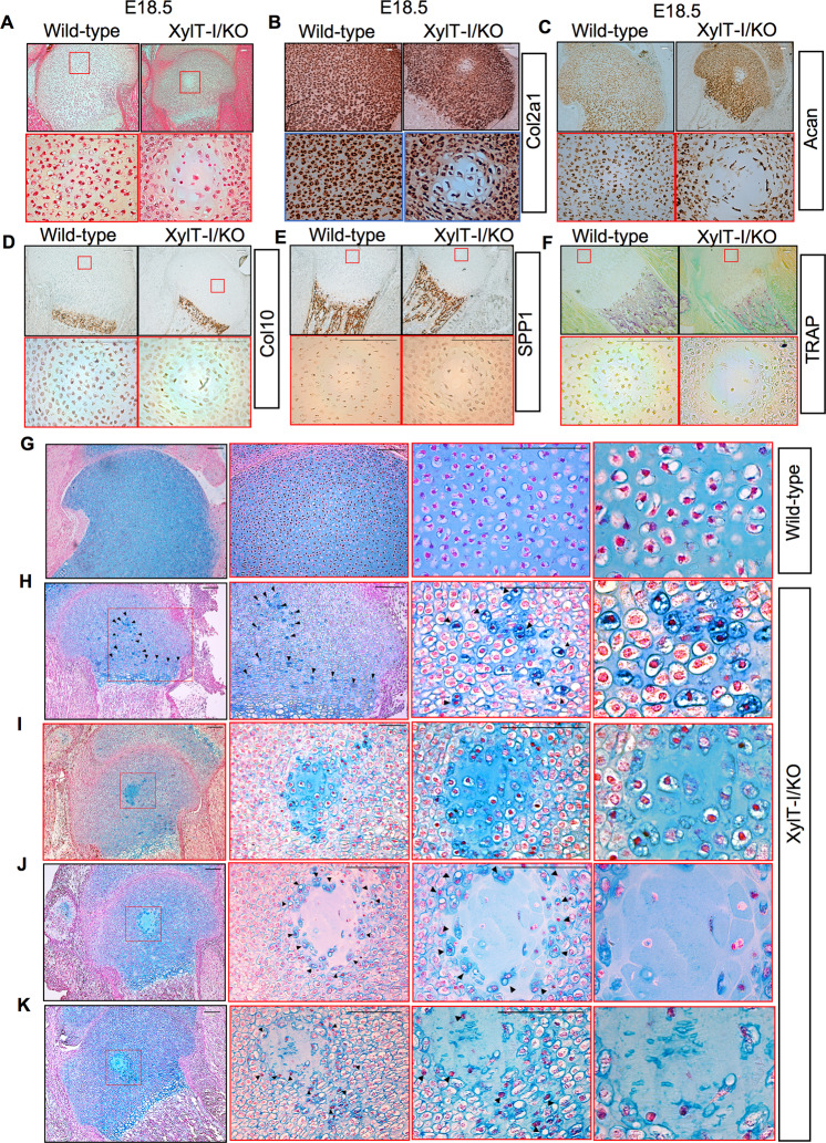
Genetic mutations in the Xylt1 gene are associated with Desbuquois dysplasia type II syndrome characterized by sever prenatal and postnatal short stature. However, the specific role of XylT-I in the growth plate is not completely understood. Here, we show that XylT-I is expressed and critical for the synthesis of proteoglycans in resting and proliferative but not in hypertrophic chondrocytes in the growth plate. We found that loss of XylT-I induces hypertrophic phenotype-like of chondrocytes associated with reduced interterritorial matrix. Mechanistically, deletion of XylT-I impairs the synthesis of long glycosaminoglycan chains leading to the formation of proteoglycans with shorter glycosaminoglycan chains. Histological and Second Harmonic Generation microscopy analysis revealed that deletion of XylT-I accelerated chondrocyte maturation and prevents chondrocytes columnar organization and arrangement in parallel of collagen fibers in the growth plate, suggesting that XylT-I controls chondrocyte maturation and matrix organization. Intriguingly, loss of XylT-I induced at embryonic stage E18.5 the migration of progenitor cells from the perichondrium next to the groove of Ranvier into the central part of epiphysis of E18.5 embryos. These cells characterized by higher expression of glycosaminoglycans exhibit circular organization then undergo hypertrophy and death creating a circular structure at the secondary ossification center location. Our study revealed an uncovered role of XylT-I in the synthesis of proteoglycans and provides evidence that the structure of glycosaminoglycan chains of proteoglycans controls chondrocyte maturation and matrix organization.


Conflict of interest statement

The authors declare no competing interests.

Figures

Fig. 1
Fig. 1. XylT-I/KO embryos display dwarfism and defects in PG synthesis.
A XylT-I/KO embryos were generated by deletion of the Exon1 of Xylt1 gene and a part of the promoter sequence resulting in a deletion of 18.5 kb gene sequence. Exons (E) are denoted by boxes and horizontal bar denote introns. B XylT-I/KO and wild-type embryos at embryonic stages E14.5, E16.5, E18.5 showing an overall reduction in body size and a frontonasal dysplasia in XylT-I/KO embryos. Graphs showing relative body and humerus length of XylT-I/KO embryos and their wild-type littermates at E14.5 and E18.5 stages, values are normalized such that control body length are set to 100%. C Sections through humerus at E18.5 stage stained with Alizarin red and Alcian blue (the length is represented by yellow arrow). D Wild-type and XylT-I/KO whole body embryos at E16.5 stained with Alizarin red and Alcian blue. XylT-I/KO embryos show frontonasal dysplasia with flat midface (yellow cercle) and skull dysplasia with narrow rib cage (red square). E In situ mRNA hybridization analysis of XylT-I expression assessed by RNAscope on sections of proximal humerus growth plate in XylT-I/KO and wild-type embryos at E18.5. F Sections of proximal humerus growth plate of wild-type and XylT-I/KO embryos at E15.5, E16.5 and E18.5 stained with Alcian blue which show strong reduction in GAGs in XylT-I/KO except in hypertrophic zone (HZ). Data are expressed as mean ± S.D. *P < 0.05, Student’s t test. Representative images from three independent experiments are shown (n = 3). Scale bar: 100 μm. G Synthesis level of PG-GAG chains in wild-type and XylT-I/KO primary chondrocytes analyzed by 35S-sulfate incorporation into GAG chains. H SDS-PAGE analysis of radiolabelled PGs and (I) GAGs chains in primary chondrocytes of wild-type and XylT-I/KO embryos. J Western blot detection of decorin from cultured medium of wild-type and XylT-I/KO primary chondrocytes. K Western blot detection of decorin from cultured medium of wild-type and XylT-I/KO primary chondrocytes following degradation of GAG-attached chains by treatment with chondroitinase ABC. Representative images from three independent experiments are shown (n = 3).
Fig. 2
Fig. 2. Increased hypertrophy and decreased interterritorial matrix in XylT-I mutant growth plate.
A HES staining of sections of humerus proximal growth plate of XylT-I/KO and wild-type embryos at E15.5 showing a shortened growth plate in XylT-I/KO embryos with larger hypertrophic zone. B Graph showing the length of RZ, PZ and HZ in XylT-I/KO embryos and their wild-type littermates at E15.5 stage. C HES staining of sections of humerus proximal growth plate of XylT-I/KO and wild-type embryos at E18.5 showing a shortened growth plate in XylT-I/KO embryos. D Graph showing the length of RZ, PZ and HZ in XylT-I/KO embryos and their wild-type littermates at E18.5 stage. EG Higher magnification of resting, proliferative and hypertrophic zone of the growth plate of wild-type and XylT-I/KO embryos at E15.5. RZ Resting zone, PZ Proliferative zone, HZ Hypertrophic zone. Data are expressed as mean ± S.D. *P < 0.05, **P < 0.01, ***P < 0.001, Student’s t test. Representative images from three independent experiments are shown (n = 3). Scale bar: 100 μm.
Fig. 3
Fig. 3. Extended hypertrophic zone and increased expression of hypertrophic and osteogenic markers in XylT-I/KO.
A Sections through humerus of wild-type and XylT-I/KO embryos at E14.5 stained with Alcian blue (humerus and hypertrophic zone length are represented by black arrow). B In situ mRNA expression analysis of Col10 at E14.5 and (C and D) of SPP1 at E14.5 and E18.5 assessed by RNAscope on sections of proximal humerus growth plate in XylT-I/KO and wild-type embryos. E Alizarin red staining of humerus sections of wild-type and XylT-I/KO embryos at E14.5 which marks mineralized tissues. FH In situ mRNA hybridization analysis of Ihh and (IK) of Runx2 at E15.5 and E18.5 assessed by RNAscope on sections of proximal humerus growth plate of XylT-I/KO and wild-type embryos (H and K higher magnification of resting and proliferative zones of the growth plate of wild-type and XylT-I/KO at E18.5). L In situ mRNA hybridization analysis of Sox9, Col2a1 and Acan assessed by RNAscope on sections of proximal humerus growth plate of XylT-I/KO and wild-type embryos at E15.5. Representative images from three independent experiments are shown (n = 3). Scale bar: 100 μm.
Fig. 4
Fig. 4. Migration of perichondrium cells to the secondary ossification center area.
A HES staining of proximal humerus growth plate sections of wild-type and XylT-I/KO embryos at E18.5. In situ mRNA hybridization analysis of (B) Col2a1, (C) Acan, (D) Col10 and (E) SPP1 in proximal humerus growth plate sections of wild-type and XylT-I/KO embryos at E18.5 using RNAscope. F TRAP staining in proximal humerus growth plate sections of wild-type and XylT-I/KO embryos at E18.5. G Alcian blue staining of the growth plate of wild-type and (H) of XylT-I/KO embryos at E18.5 showing the presence in XylT-I/KO of a group of cells with condensed nuclei and strongly stained with Alcian blue migrating from the perichondrium and invading the growth plate until reaching the secondary ossification center area. I These cells form a circular structure and (J) became hypertrophic (K) then degraded. Representative images from three independent experiments are shown (n = 3). Scale bar: 100 μm.
Fig. 5
Fig. 5. Upregulation of FGFR3 and activation of ERK1/2 in XylT-I/KO embryos.
A Immunohistochemistry of phospho-ERK1/2 in sections of humerus of wild-type and XylT-I/KO embryos at E18.5. B In situ mRNA hybridization of FGFR3 in the growth plate of wild-type and XylT-I/KO embryos at E18.5 assessed by RNAscope. Representative images from three independent experiments are shown (n = 3). Scale bar: 100 μm.
Fig. 6
Fig. 6. Reduced body and elements length and impaired trabecular and cortical bone formation in XylT-I/cKO mice.
A XylT1flox/flox mice with loxP sites flanking exon 5 were crossed with Col2α1-CreERTM mice carrying tamoxifen-inducible Col2a1-promoter driven Cre recombinase to generate XylT1flox/flox; Col2α1-CreERTM mice. Exon 5 is deleted following tamoxifen administration to generate XylT-I/cKO mice. B The body size of XylT-I/cKO mice at 8 weeks after birth shows 20% reduction in length with shortened snouts, limbs, and tails compared with control littermates. C XylT-I/cKO mice display reduction in femurs and tibias length. Values are normalized such that control femurs and tibias are set to 100%. Whole mount skeletal staining with Alcian blue and Alizarin red showing dwarfism phenotype characterized by shortened axial and appendicular skeletons, shortening of the skull, smaller rib cage, shortening humerus and early ossification of trachea. D Micro-CT scan analysis of trabecular bone in XylT-I/cKO and wild-type mice. Graph showing several measurements of trabecular and cortical bone parameters including bone volume to total volume fraction (BV/TV), trabecular number (Tb.N), trabecular thickness (Tb.Th), trabecular separation (Tb.Sp), trabecular pattern factor (Tb.Pf), cortical thickness (Ct.Th), structure model index (SMI), tissue mineral density (TMD) and trabecular bone density (BMD). Values are normalized such that control is defined as 1. E Micro-CT scan analysis showing larger femoral epiphysis, more developed and more ossified femoral head (white squares) and thicker cortical bone in XylT-I/cKO mice, compared with control mice. F TRAP staining of tibial cancellous and cortical bone in XylT-I/cKO mice, compared with control mice. G RT-qPCR analysis of the expression of bone formation markers and bone transcriptions factors in cortical bone in XylT-I/cKO mice, compared with control mice. Q-PCR values were normalized for the housekeeping gene ribosomal protein S29. Data are expressed as mean ± S.D. *P < 0.05, **P < 0.01, ***P < 0.001, Student’s t test. Representative images from three independent experiments are shown (n = 3). Scale bar: 100 μm.
Fig. 7
Fig. 7. Disruption in chondrocytes and collagen fibers organization and accelerated maturation of chondrocytes in the growth plate of XylT-I/cKO mice.
A HES staining of tibias sections of 10 days old XylT-I/cKO and control mice showing severe reduction of the hypertrophic zone in XylT-I/cKO mice. B Alcian blue staining of PGs revealed significantly decreased in GAG content in the resting and proliferative zones but not in hypertrophic zone in XylT-I/cKO mice, compared to control mice. C HES and Alcian blue staining of tibias growth plate of 4 weeks old XylT-I/cKO and control mice show disruption of chondrocytes columnar organization and reduced PGs content in XylT-I/cKO mice. D Second Harmonic Generation (SHG) imaging assessment of collagen fibers in the growth plate of 4 weeks old XylT-I/cKO and control mice showing collagen fibers run parallel to each other in control mice but disorganized and oriented in multiple directions in XylT-I/cKO mice. Autofluorescence (grey) and SHG signal (green) indicates the collagen in the tissue. E In situ mRNA expression analysis showing increased expression of the chondrogenic markers Acan and Col2α1, and of hypertrophic marker Col10 in 10 days old XylT-I/cKO mice compared to wild-type mice. F In situ mRNA expression analysis showing increased expression of Spp1, β-Glap and Ihh and (G) of the terminal hypertrophic differentiation marker MMP13 in the growth plate of 10 days old XylT-I/cKO mice, compared with control mice. RZ Resting zone, PZ Proliferative zone, PhZ Prehypertrophic zone, HZ Hypertrophic zone. Representative images from three independent experiments are shown (n = 3). Scale bar: 100 μm.

Similar articles

Cited by

References

    1. Muir H. Proteoglycans of cartilage. J Clin Pathol Suppl. 1978;12:67–81. doi: 10.1136/jcp.31.Suppl_12.67. - DOI - PMC - PubMed
    1. Kjellen L, Lindahl U. Proteoglycans: structures and interactions. Annu Rev Biochem. 1991;60:443–75. doi: 10.1146/annurev.bi.60.070191.002303. - DOI - PubMed
    1. Couchman JR. Transmembrane signaling proteoglycans. Annu Rev Cell Dev Biol. 2010;26:89–114. doi: 10.1146/annurev-cellbio-100109-104126. - DOI - PubMed
    1. Iozzo RV, Schaefer L. Proteoglycan form and function: a comprehensive nomenclature of proteoglycans. Matrix Biol. 2015;42:11–55. doi: 10.1016/j.matbio.2015.02.003. - DOI - PMC - PubMed
    1. Melrose J, Shu C, Whitelock JM, Lord MS. The cartilage extracellular matrix as a transient developmental scaffold for growth plate maturation. Matrix Biol. 2016;52-54:363–83. doi: 10.1016/j.matbio.2016.01.008. - DOI - PubMed

Publication types

LinkOut - more resources