

Systematic detection of tertiary structural modules in large RNAs and RNP interfaces by Tb-seq
- PMID: 37296103
- PMCID: PMC10255950
- DOI: 10.1038/s41467-023-38623-1
Systematic detection of tertiary structural modules in large RNAs and RNP interfaces by Tb-seq
Abstract
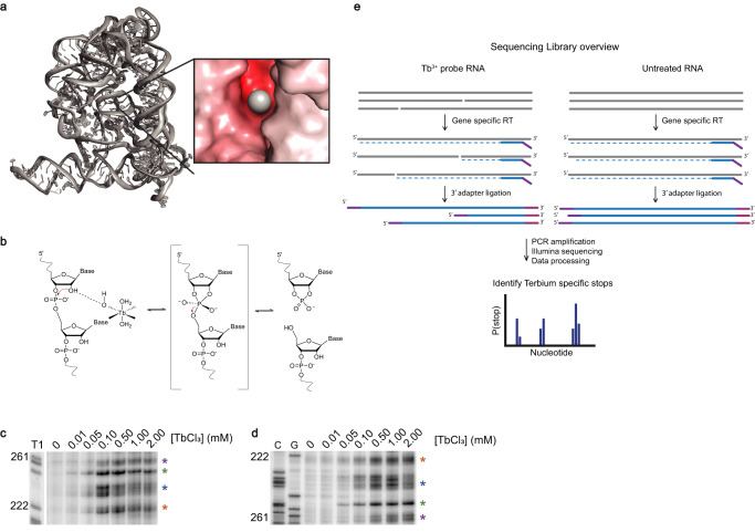
Compact RNA structural motifs control many aspects of gene expression, but we lack methods for finding these structures in the vast expanse of multi-kilobase RNAs. To adopt specific 3-D shapes, many RNA modules must compress their RNA backbones together, bringing negatively charged phosphates into close proximity. This is often accomplished by recruiting multivalent cations (usually Mg2+), which stabilize these sites and neutralize regions of local negative charge. Coordinated lanthanide ions, such as terbium (III) (Tb3+), can also be recruited to these sites, where they induce efficient RNA cleavage, thereby revealing compact RNA 3-D modules. Until now, Tb3+ cleavage sites were monitored via low-throughput biochemical methods only applicable to small RNAs. Here we present Tb-seq, a high-throughput sequencing method for detecting compact tertiary structures in large RNAs. Tb-seq detects sharp backbone turns found in RNA tertiary structures and RNP interfaces, providing a way to scan transcriptomes for stable structural modules and potential riboregulatory motifs.
© 2023. The Author(s).
Conflict of interest statement
A patent application on MarathonRT has been filed by Yale University. Yale university has submitted a patent application pertaining to work outlined in this study. Inventors include A.M.P, S.P and M.D.S. C.B.W is a consultant for Exscientia. The remaining authors declare no competing interests.
Figures

Similar articles
-
Terbium-mediated footprinting probes a catalytic conformational switch in the antigenomic hepatitis delta virus ribozyme.J Mol Biol. 2004 Aug 6;341(2):389-403. doi: 10.1016/j.jmb.2004.05.074. J Mol Biol. 2004. PMID: 15276831
-
Probing non-selective cation binding in the hairpin ribozyme with Tb(III).J Mol Biol. 2000 May 5;298(3):539-55. doi: 10.1006/jmbi.2000.3691. J Mol Biol. 2000. PMID: 10772868
-
Monitoring global structural changes and specific metal-ion-binding sites in RNA by in-line probing and Tb(III) cleavage.Methods Mol Biol. 2014;1086:143-58. doi: 10.1007/978-1-62703-667-2_8. Methods Mol Biol. 2014. PMID: 24136602
-
Probing RNA structure and metal-binding sites using terbium(III) footprinting.Curr Protoc Nucleic Acid Chem. 2003 Aug;Chapter 6:Unit 6.8. doi: 10.1002/0471142700.nc0608s13. Curr Protoc Nucleic Acid Chem. 2003. PMID: 18428913 Review.
-
Folding of RNA tertiary structure: Linkages between backbone phosphates, ions, and water.Biopolymers. 2013 Dec;99(12):1105-13. doi: 10.1002/bip.22249. Biopolymers. 2013. PMID: 23568785 Free PMC article. Review.
Cited by
-
Characterization and implementation of the MarathonRT template-switching reaction to expand the capabilities of RNA-seq.RNA. 2024 Oct 16;30(11):1495-1512. doi: 10.1261/rna.080032.124. RNA. 2024. PMID: 39174298 Free PMC article.
-
Long Non-Coding RNAs as "MYC Facilitators".Pathophysiology. 2023 Sep 1;30(3):389-399. doi: 10.3390/pathophysiology30030030. Pathophysiology. 2023. PMID: 37755396 Free PMC article. Review.
-
Limits of experimental evidence in RNA secondary structure prediction.Front Bioinform. 2024 Feb 22;4:1346779. doi: 10.3389/fbinf.2024.1346779. eCollection 2024. Front Bioinform. 2024. PMID: 38456157 Free PMC article. No abstract available.
References
Publication types
MeSH terms
Substances
Grants and funding
LinkOut - more resources
Full Text Sources
Other Literature Sources
