. 2023 Jun 9;14(6):356.
doi: 10.1038/s41419-023-05868-z.

HNF4A-BAP31-VDAC1 axis synchronously regulates cell proliferation and ferroptosis in gastric cancer

Affiliations

HNF4A-BAP31-VDAC1 axis synchronously regulates cell proliferation and ferroptosis in gastric cancer

Qingqing Zhou et al. Cell Death Dis. .

Abstract

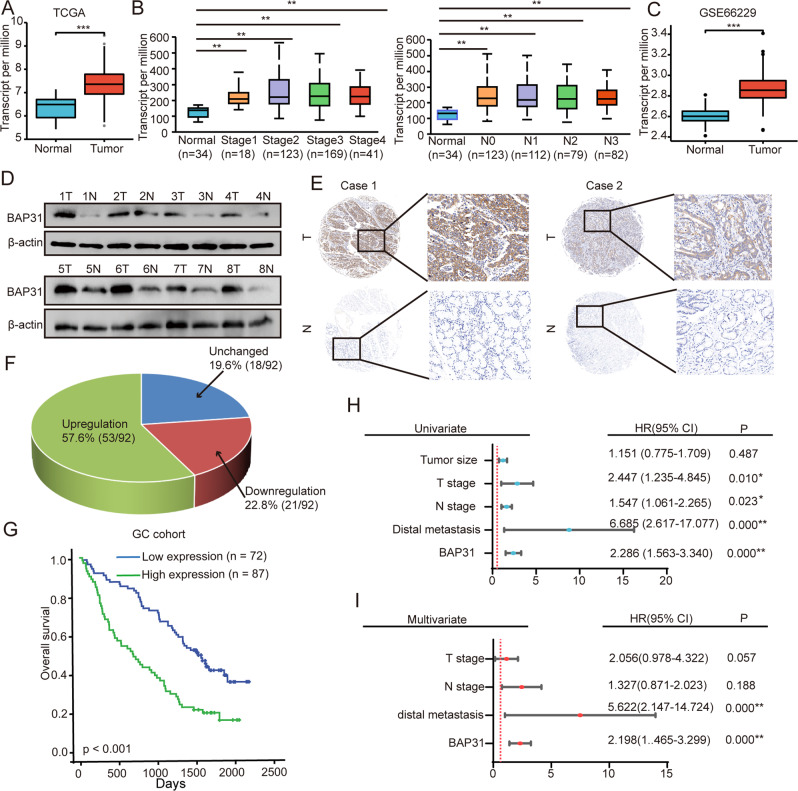
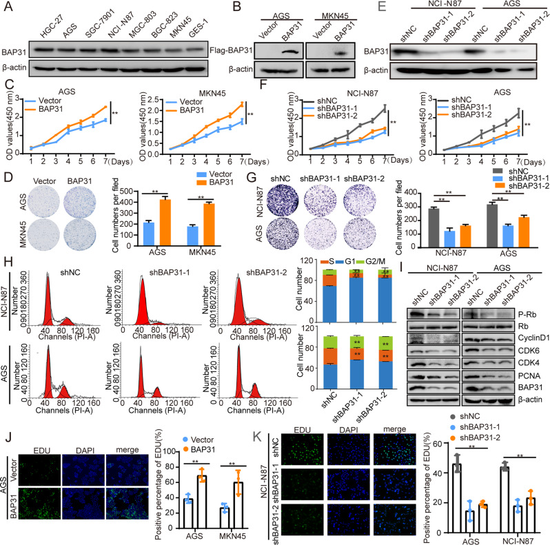
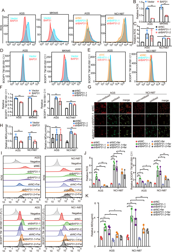
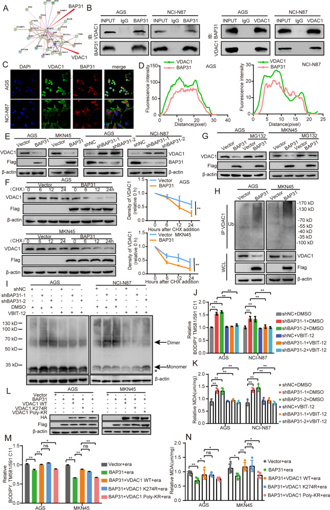
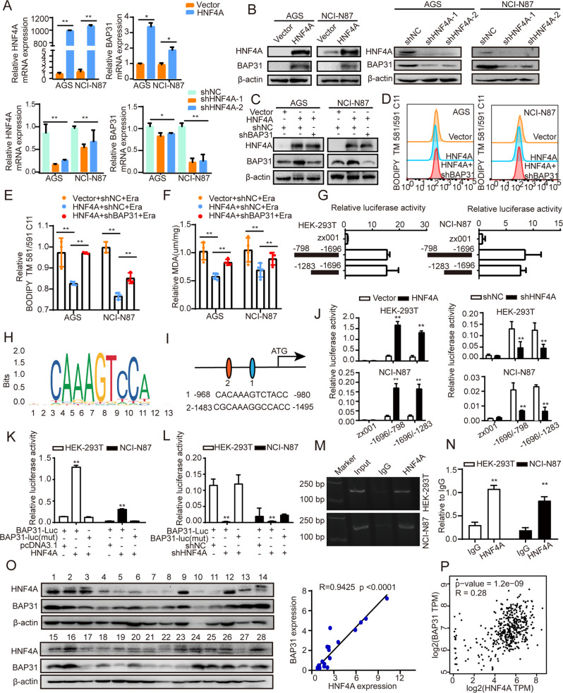
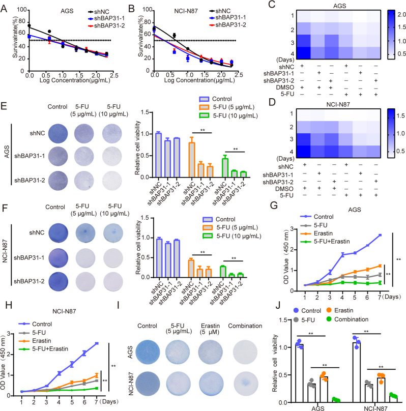
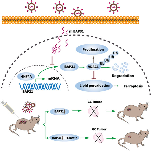
B cell receptor associated protein 31 (BAP31) is closely associated with tumor progression, while the role and mechanism of BAP31 in gastric cancer (GC) remains unknown. This study explored that BAP31 was upregulated in GC tissues and high expression indicated poor survival of GC patients. BAP31 knockdown inhibited cell growth and induced G1/S arrest. Moreover, BAP31 attenuation increased the lipid peroxidation level of the membrane and facilitated cellular ferroptosis. Mechanistically, BAP31 regulated cell proliferation and ferroptosis by directly binding to VDAC1 and affected VDAC1 oligomerization and polyubiquitination. HNF4A was bound to BAP31 at the promoter and increased its transcription. Furthermore, knockdown of BAP31 inclined to make GC cells vulnerable to 5-FU and ferroptosis inducer, erastin, in vivo and in vitro. Our work suggests that BAP31 may serve as prognostic factor for gastric cancer and act as potential therapeutic strategy for gastric cancer.


Conflict of interest statement

The authors declare no competing interests.

Figures

Fig. 1
Fig. 1. GC with increased BAP31 expression indicate a worse prognosis.
A BAP31 was analyzed in GC tissues and in non-cancerous gastric tissues derived from TCGA data. B TCGA datasets were used to analyze the expression of BAP31 in different GC stages, and nodal metastasis. C BAP31 expression in GC tissues as well as noncancerous gastric tissues was analyzed using the GSE66229 dataset. D An analysis of BAP31 expression in GC tissues compared to noncancerous gastric tissues by western blot. E Representative IHC images showing the level of BAP31 in GC tissues and the corresponding noncancerous tissues. F Pie chart illustrating the proportion of upregulation, unchanged and downregulation in BAP31 for comparison between GC tissues and noncancerous tissues. G Kaplan–Meier analysis of overall survival in GC patients with differential BAP31 expression in 159 samples. Univariate (H) and multivariate (I) Cox proportional hazard analyses were conducted to evaluate the HR of BAP31 in GC. *p < 0.05, **p < 0.01.
Fig. 2
Fig. 2. BAP31 promotes GC cell growth.
A Western blot was applied to determine the endogenous level of BAP31 in GC cell lines. B Western blot was applied to detect the expression of BAP31 in GC cells with BAP31 overexpression. C CCK8 assay was utilized to evaluate the influence of BAP31 overexpression on GC cell growth. D Colony formation assay was used to evaluate the influence of BAP31 overexpression on GC cell growth. E Western blot was applied to detect the expression of BAP31 in GC cells with BAP31 knockdown. F CCK8 assay was utilized to evaluate the influence of BAP31 knockdown on GC cell growth. G Colony formation assay was used to evaluate the influence of BAP31 knockdown on GC cell growth. H Cell cycle distributions in BAP31 knockdown GC cells were detected by flow cytometry. I Western blot was applied to detect the cell cycle-related proteins with BAP31 knockdown GC cells. EdU assay was used to assess growth on GC cells with BAP31 overexpression (J) or BAP31 knockdown (K). *p < 0.05, **p < 0.01.
Fig. 3
Fig. 3. BAP31 attenuation facilitates lipid peroxidation and ferroptosis.
AC Flow cytometry was applied to detect ROS fluorescence on GC cells under BAP31 overexpression or knockdown, and cells were pre-treated with erastin (5 μM) for 12 h. DF The GC cells with BAP31 overexpression or BAP31 knockdown pretreated with erastin (5 μM) for 12 h, and lipid ROS production was tested. G Confocal imaging detected lipid peroxidation under BAP31 knockdown in GC cells pretreated with erastin (5 μM) for 12 h. H BAP31-overexpressing or BAP31-knockdown GC cells pretreated with erastin (5 μM) for 12 h, and MDA content was assessed. I, J BAP31-knockdown GC cells pretreated with erastin (5 µM) for 12 h in the presence or absence of 2 μmol/L Fer-1, and then lipid ROS production was observed. K GC cells pretreated with erastin (5 µM) for 12 h in the presence or absence of 2 μmol/L Fer-1, and MDA content was assayed. *p < 0.05, **p < 0.01.
Fig. 4
Fig. 4. Knockdown of BAP31 sensitizes GC cells to ferroptosis inducer, erastin.
A The IC50s of erastin were analyzed in AGS and NCI-N87 cells with BAP31 knockdown. B Heatmap demonstrated concerted reaction to 5 μM erastin combined with BAP31 attenuation in GC cells. C Clone formation assays exhibited concerted reaction to erastin (5 μM, 10 μM) combined with BAP31 attenuation in GC cells. D Subcutaneous xenograft model and injection schedule were formulated in nude mice. NCI-N87 cells transfected with shNC or shBAP31 were subcutaneously injected into the flanks of nude mice administrated without or with erastin, and representative images of dissected tumors (E), tumor weight (F) and tumor growth curves (G) of mice. H HE and IHC assay for Ki-67 and PTGS2 were performed in isolated tumor tissues. I Body weight was measured in the above-mentioned mice. *p < 0.05, **p < 0.01.
Fig. 5
Fig. 5. BAP31 interact with VDAC1 and affect its protein stability.
A STRING database was utilized to predict proteins that interact with BAP31. B Immunoprecipitation was conducted to examine the relationship between BAP31 and VDAC1. C, D The interaction between BAP31 and VDAC1 were observed through immunofluorescence assays. E Western blot was used to assess the protein level of VDAC1 in BAP31-overexpressed or BAP31-knockdown GC cells. F BAP31 overexpression group and control group were pretreated with cycloheximide (CHX), then the protein level of VDAC1 was detected. G BAP31 overexpression group and control group were pretreated with MG132 6 h, then the protein level of VDAC1 was detected. H The ubiquitination of VDAC1 in BAP31 overexpression group and control group. I We attenuated BAP31 in AGS and NCI-N87, and treated with DMSO or VBIT-12 respectively. Then, cells in each group were harvested and incubated in the presence of ethylene glycol bis (succinimidyl succinate) to cross-link proteins and then subject to western blot to assess the oligomeric status of VDAC1. Arrows indicate monomer and dimer forms of VDAC1. Asterisk indicates the intramolecular cross-linked bands. BAP31 knockdown GC cells pretreated with erastin (5 µM) for 12 h in the presence or absence of 10 µM VBIT-12 (VDAC1 oligomerization inhibitor), where lipid ROS production (J) and MDA content (K) were assayed. L The expression of BAP31 and VDAC1 was detected in BAP31-overexpressing cells transfected with VDAC1 WT or two ubiquitination mutants (VDAC1 K274R and Poly-KR). BAP31-overexpressing GC cells transfected with VDAC1 WT or two ubiquitination mutants treated with erastin (5 µM) for 12 h, where lipid ROS production (M) and MDA content (N) were tested. ns.p > 0.05, *p < 0.05, **p < 0.01.
Fig. 6
Fig. 6. HNF4A binds at BAP31 promoter and augments BAP31 transcription.
A, B The mRNA and protein level of BAP31 were detected in GC cells with HNF4A overexpression or knockdown. C The level of HNF4A and BAP31 was detected in HNF4A-overexpressing cells transfected with BAP31 shRNA. DF Lipid ROS and MDA content were assayed in HNF4A-overexpressing cells treated with BAP31 shRNA treated with erastin (5 µM) for 12 h. G Relative luciferase activities were assessed in HEK-293T and NCI-N87 cells transfected with different truncations of BAP31 promoter. H HNF4A binding motif was predicted in JASPAR. I JASPAR analysis revealed two potential HNF4A-binding sites (scores >10) within the promoter region of BAP31. J HEK-293T and NCI-N87 cells transfected with different truncations of BAP31 promoter, in the presence of HNF4A overexpression or knockdown respectively, then relative luciferase activities were assessed. K, L Relative luciferase activities were assessed in HEK-293T and NCI-N87 cells treated with BAP31 luciferase reporter vectors (wild-type or mutant in HNF4A-binding sites, -1495/-1483 bp), in the presence of HNF4A overexpression or knockdown, respectively. M Agarose electrophoresis for ChIP analysis of HNF4A binding at BAP31 promoter. N ChIP-qPCR analysis of HNF4A binding at BAP31 promoter. O Western blot analysis of BAP31 and HNF4A expression in GC tissues (n = 28). P The relationship between HNF4A and BAP31 in GC tissues was analyzed using TCGA datasets (R = 0.28, p < 0.001). *p < 0.05, **p < 0.01.
Fig. 7
Fig. 7. Knockdown of BAP31 sensitizes GC cells to 5-FU and combining 5-FU with erastin amplifies anti-tumor effects.
A, B The IC50s of 5-FU in AGS and NCI-N87 cells with BAP31 knockdown were analyzed. C, D Heatmap demonstrated synergistic response to 5 μg/ml 5-FU combined with BAP31 knockdown in AGS and NCI-N87 cells. E, F Clone formation assays showed synergistic response to 5-FU (5 μg/ml, 10 μg/ml) combined with BAP31 knockdown in AGS and NCI-N87 cells. GJ CCK8 and clone formation assays showed combination of erastin with 5-FU treatment exhibited higher inhibitory effects on cell growth compared with mono treatment in AGS and NCI-N87. *p < 0.05, **p < 0.01.
Fig. 8
Fig. 8. Schematic representation of HNF4A-BAP31-VDAC1 axis that synchronously regulates proliferation and ferroptosis in gastric cancer.
HNF4A directly binds to BAP31 promoter and augments its transcription. BAP31 promotes cell proliferation and inhibits lipid peroxidation and ferroptosis. BAP31 overexpression directly induces VDAC1 protein degradation via the ubiquitin-proteasome pathway to promote GC progression. The shBAP31 impiars BAP31 translation and enhances the antitumor effects of ferroptosis inducer, erastin, which may serve as promising therapeutic strategy in anti -tumor treatment.

Similar articles

Cited by

References

    1. Smyth EC, Nilsson M, Grabsch HI, van Grieken NC, Lordick F. Gastric cancer. Lancet. 2020;396:635–48. doi: 10.1016/S0140-6736(20)31288-5. - DOI - PubMed
    1. Siegel RL, Miller KD, Fuchs HE, Jemal A. Cancer statistics, 2021. CA Cancer J Clin. 2021;71:7–33. doi: 10.3322/caac.21654. - DOI - PubMed
    1. Petryszyn P, Chapelle N, Matysiak-Budnik T. Gastric cancer: where are we heading? Dig Dis. 2020;38:280–5. doi: 10.1159/000506509. - DOI - PubMed
    1. Machlowska J, Baj J, Sitarz M, Maciejewski R, Sitarz R. Gastric cancer: epidemiology, risk factors, classification, genomic characteristics and treatment strategies. Int J Mol Sci. 2020;21:4012. doi: 10.3390/ijms21114012. - DOI - PMC - PubMed
    1. Kim KM, Adachi T, Nielsen PJ, Terashima M, Lamers MC, Köhler G, et al. Two new proteins preferentially associated with membrane immunoglobulin D. EMBO J. 1994;13:3793–3800. doi: 10.1002/j.1460-2075.1994.tb06690.x. - DOI - PMC - PubMed

Publication types

MeSH terms

Substances