. 2023 Jun 9;14(1):3416.
doi: 10.1038/s41467-023-39140-x.

A 2.2 Å cryoEM structure of a quinol-dependent NO Reductase shows close similarity to respiratory oxidases

Affiliations

A 2.2 Å cryoEM structure of a quinol-dependent NO Reductase shows close similarity to respiratory oxidases

Alex J Flynn et al. Nat Commun. .

Abstract

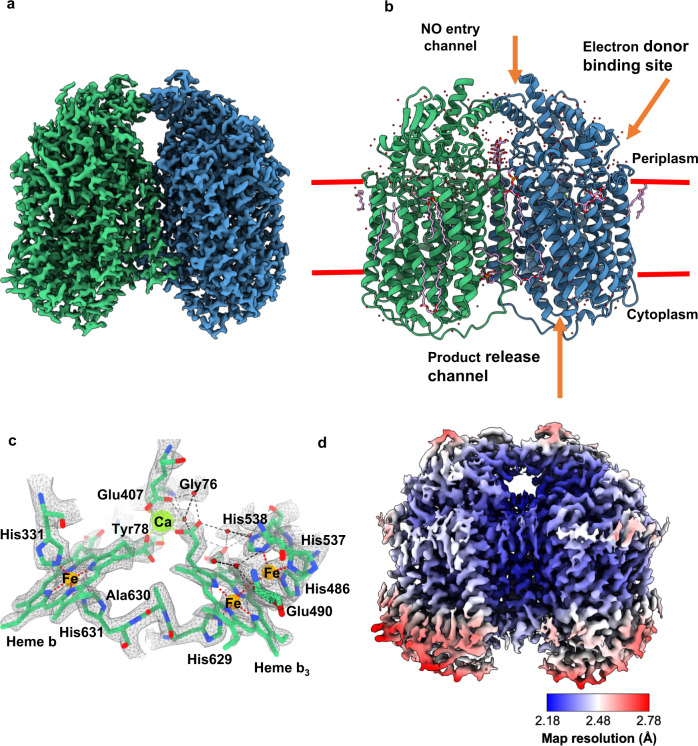
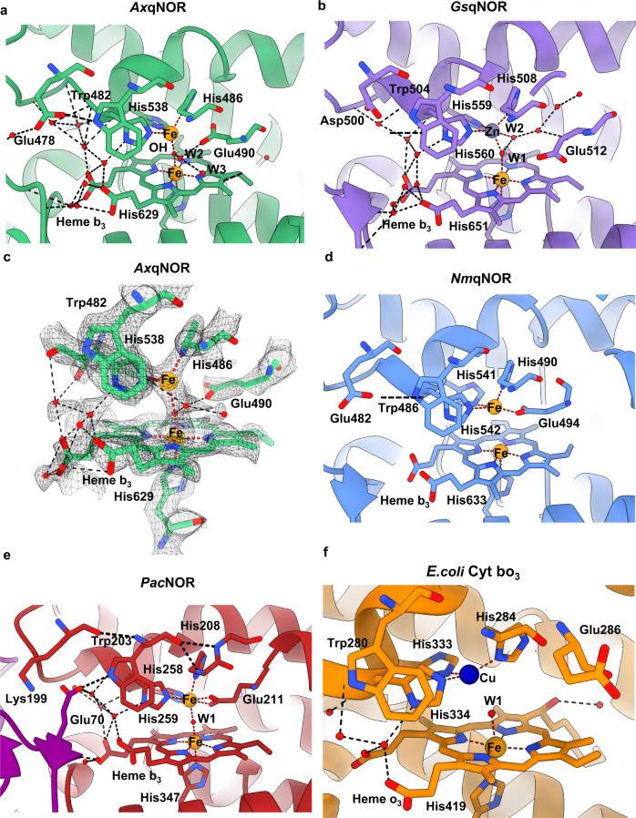
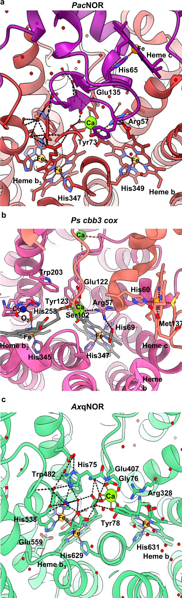
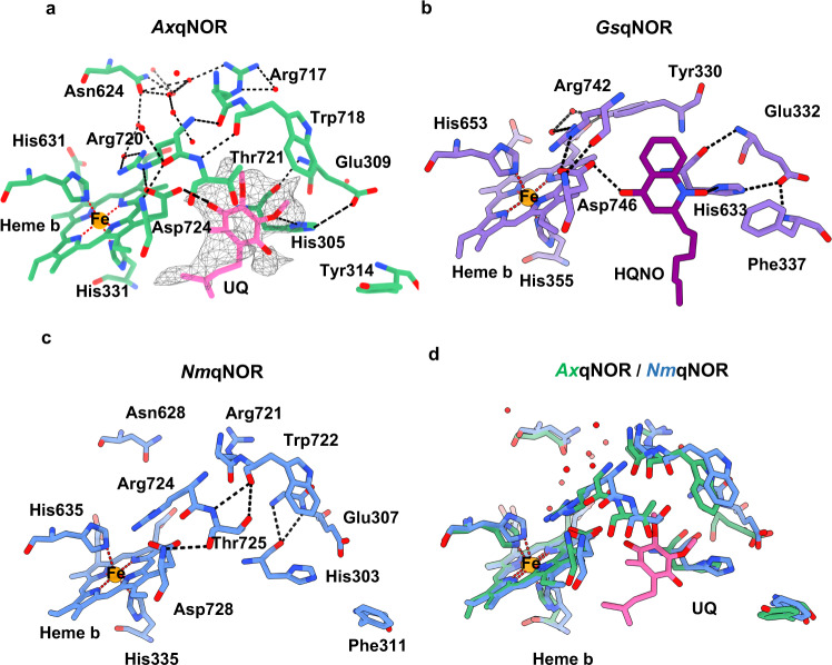
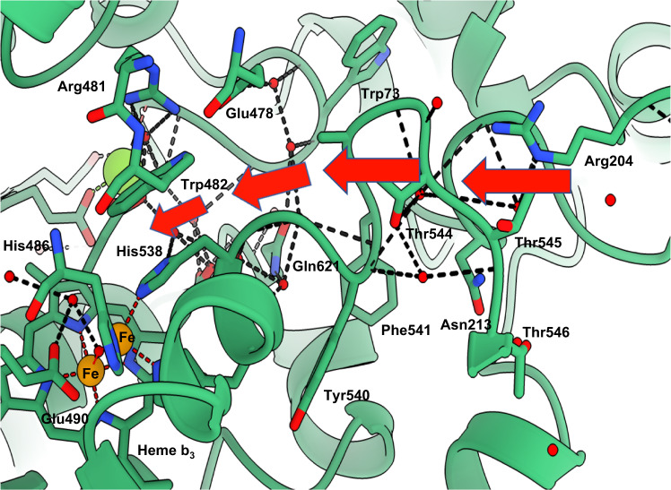
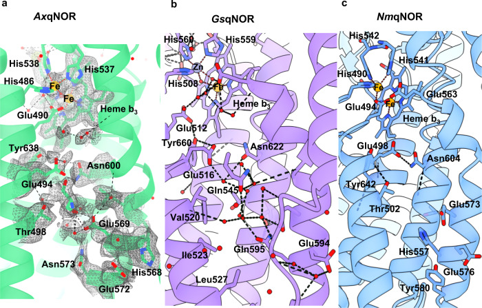
Quinol-dependent nitric oxide reductases (qNORs) are considered members of the respiratory heme-copper oxidase superfamily, are unique to bacteria, and are commonly found in pathogenic bacteria where they play a role in combating the host immune response. qNORs are also essential enzymes in the denitrification pathway, catalysing the reduction of nitric oxide to nitrous oxide. Here, we determine a 2.2 Å cryoEM structure of qNOR from Alcaligenes xylosoxidans, an opportunistic pathogen and a denitrifying bacterium of importance in the nitrogen cycle. This high-resolution structure provides insight into electron, substrate, and proton pathways, and provides evidence that the quinol binding site not only contains the conserved His and Asp residues but also possesses a critical Arg (Arg720) observed in cytochrome bo3, a respiratory quinol oxidase.


Conflict of interest statement

The authors declare no competing interests.

Figures

Fig. 1
Fig. 1. The structure of AxqNOR and its new features identified in a high-resolution cryoEM map.
a cryoEM map for the measured Coulomb potential of the scattered atoms of AxqNOR determined by single-particle cryoEM to 2.2 Å resolution. Map colored by chain (A in green and B in blue). b Dimeric AxqNOR in the plane of the lipid bilayer (red lines) colored by chain (A in green and B in blue), with waters colored as red spheres, DTM (decyl-thio-maltoside) detergent and LOP (lauryl oleyl phosphatidyl-ethanolamine (LOP) as magenta sticks. c AxqNOR catalytic core formed of two adjacent heme groups linked by a calcium ion (green sphere) shown within the cryoEM density map. Iron ions are shown as orange spheres, water molecules as red spheres, and protein and heme groups as green sticks. All density maps contoured to a sigma level of 0.034 except for that corresponding to water molecules and Glu490, which is contoured to 0.020. d Map of AxqNOR cryoEM density colored by local resolution.
Fig. 2
Fig. 2. Details of high-resolution AxqNOR and its comparison with other family members GsqNOR, NmqNOR, PacNOR, and E. coli cytochrome bo3.
a Binuclear center of AxqNOR with FeB coordinated by His486, His537, and His538 and µ-oxo bridge O. Iron atoms and water molecules shown as orange and red spheres, respectively. Hydrogen bonds and metal coordinating bonds are shown as black and red dashed lines, respectively. This scheme is used throughout the figure. b Binuclear center of GsqNOR (PDB ID: 3AYG) with a penta-coordinated zinc center. Glu512 is not bonded to ligands of the non-heme iron center. Zn is shown as a gray sphere. c Binuclear center of AxqNOR with the density map contoured to a sigma level of 0.034, except for that corresponding to Glu490 and water molecules which is contoured at 0.020. d Binuclear center of NmqNOR (PDB ID: 6L3H) with a non-heme FeB, which is coordinated by three histidine residues and Glu494. Fe ions are shown as orange spheres. e Binuclear center of PacNOR (PDB: 3O0R) with a penta-coordinated FeB center. Glu211 is bonded to a non-heme iron center. Water molecule W1 is bound to FeB and heme b3 Fe. f Binuclear center of E. coli cytochrome bo3 (PDB: 7N9Z) with a tri-coordinated Cu center. Cu is shown as a blue sphere.
Fig. 3
Fig. 3. Comparison of AxqNOR, PacNOR, and Ps cbb3 cox calcium-binding sites.
a The PacNOR Ca binding site is located on the interface between the CytC domain (purple) and the cNOR domain (red). Residues proposed to be involved in the electron transfer from heme c to heme b and heme bh are shown as sticks (PDB ID 3O0R). Water molecules are shown as red spheres, Fe atoms are shown as orange spheres, and calcium ions as green spheres. Hydrogen bonds are shown as black dashed lines and Ca and Fe coordination bonds are shown in green/red dotted lines, respectively. This scheme is used in the figure. b Ps cbb3 cox Ca binding site (PDB: 5DJQ), Ca ion with hemisphere coordination is responsible for non-covalent fixation of ring D carboxylates of heme b and heme b3 from subunit N (pink) it also has a role of supporting subunit O (salmon) linker via Ser102-OD and Glu122-OE2. Cu atom is shown as a dark blue sphere, O2 molecule, bound between heme b3 Fe and CuB is shown as a red stick. c AxqNOR Ca binding site, with protein shown as green ribbons and key residues highlighted as sticks.
Fig. 4
Fig. 4. Potential electron donor binding site identified in AxqNOR map.
a Proposed electron donor binding site in AxqNOR showing weak cryoEM density for ubiquinol (UQ). AxqNOR is colored green with residues lining the site highlighted as sticks, and the ubiquinol molecule is shown as pink sticks. The cryoEM density is contoured to a sigma level of 0.01 and shown as gray mesh. Water molecules and Fe ions are shown in red and orange spheres, respectively. This scheme is used throughout the figure. b HQNO binding site in GsqNOR (PDB: 3AYG). GsqNOR is colored in purple, with residues lining the binding site highlighted as sticks, HQNO molecule is colored in dark purple. c Potential ubiquinol binding site in NmqNOR (PDB: 6L3H). NmqNOR is colored in blue, with residues lining the binding site highlighted as sticks. d Superposition of AxqNOR (green) and NmqNOR (blue) and the potential UQ binding site.
Fig. 5
Fig. 5. AxqNOR putative NO path indicated by red arrows.
Cartoon representation of the molecule around putative NO path, indicated by red arrows. The path starts in the periplasmic part of AxqNOR and contains many charged and conserved residues. Important residues are shown as sticks, water molecules, iron, and Ca ions are represented by red, orange, and green spheres, respectively. Coordinating bonds are indicated by red dashed lines and hydrogen bonds by black lines. Labeled residues are fully conserved, except for Trp73 and Phe541.
Fig. 6
Fig. 6. Comparison of product release channels in structurally characterized qNORs.
a Product release path of qNOR from Alcaligenes xylosoxidans (AxqNOR) from the binuclear center (top) to the cytoplasmic side (bottom). AxqNOR colored in green with residues lining the proposed path in AxqNOR shown as sticks. Fe atoms and water molecules are shown as orange and red spheres, respectively. Hydrogen bonds and metal coordination bonds are shown as black and red dashed lines, respectively. This scheme is used throughout the figure. b Product release channel in qNOR from Geobacillus stearothermophilus (GsqNOR) (PDB ID: 3AYG) colored in purple, with residues lining its proposed proton channel highlighted as sticks. Zn is shown as a gray sphere. c Product release channel in qNOR from Neisseria meningitidis (NmqNOR) colored in blue, with residues lining its proposed proton channel highlighted as sticks (PDB ID: 6L3H).
Fig. 7
Fig. 7. Ordered lipids and detergents present in the cryoEM map of AxqNOR.
a Overview of additional non-protein density in the AxqNOR map. Chain A density is shown in green, chain B density is shown in blue, LOP lipid density is shown in orange, and DTM detergent is shown in salmon. The density for protein is contoured to a sigma level of 0.030 and the density for detergent and lipids is contoured to a sigma level of 0.008. This scheme is used throughout the figure. b Modeled LOP lipids found at the protein exterior and dimer interface. c Modeled fragment of LOP crossing the dimer interface and protruding into the product release channel. d DTM detergent at the dimer interface.

References

    1. Gonska N, et al. Characterization of the quinol-dependent nitric oxide reductase from the pathogen Neisseria meningitidis, an electrogenic enzyme. Sci. Rep. 2018;8:1–13. doi: 10.1038/s41598-018-21804-0. - DOI - PMC - PubMed
    1. Al-Attar S, De Vries S. An electrogenic nitric oxide reductase. FEBS Lett. 2015;589:2050–2057. doi: 10.1016/j.febslet.2015.06.033. - DOI - PubMed
    1. Hendriks JHM, Jasaitis A, Saraste M, Verkhovsky MI. Proton and electron pathways in the bacterial nitric oxide reductase. Biochemistry. 2002;41:2331–2340. doi: 10.1021/bi0121050. - DOI - PubMed
    1. Anjum MF, Stevanin TM, Read RC, Moir JWB. Nitric oxide metabolism in Neisseria meningitidis. J. Bacteriol. 2002;184:2987–2993. doi: 10.1128/JB.184.11.2987-2993.2002. - DOI - PMC - PubMed
    1. Rock JD, Moir JWB. Microaerobic denitrification in Neisseria meningitidis. Biochem. Soc. Trans. 2005;33:134–136. doi: 10.1042/BST0330134. - DOI - PubMed

Publication types

MeSH terms