. 2023 Jun 9;14(1):3386.
doi: 10.1038/s41467-023-39135-8.

In vivo PAR-CLIP (viP-CLIP) of liver TIAL1 unveils targets regulating cholesterol synthesis and secretion

Affiliations

In vivo PAR-CLIP (viP-CLIP) of liver TIAL1 unveils targets regulating cholesterol synthesis and secretion

Hasan Vatandaslar et al. Nat Commun. .

Abstract

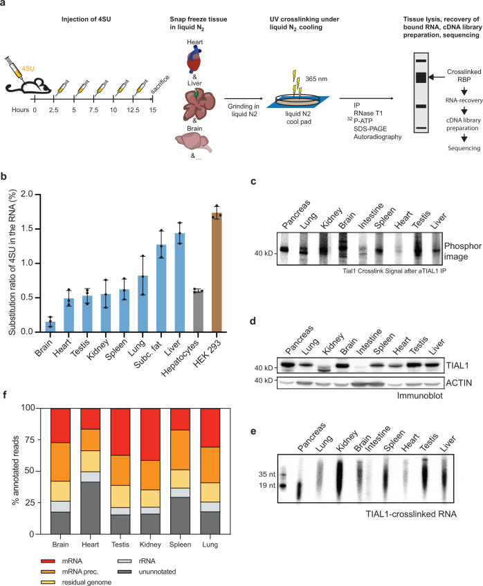
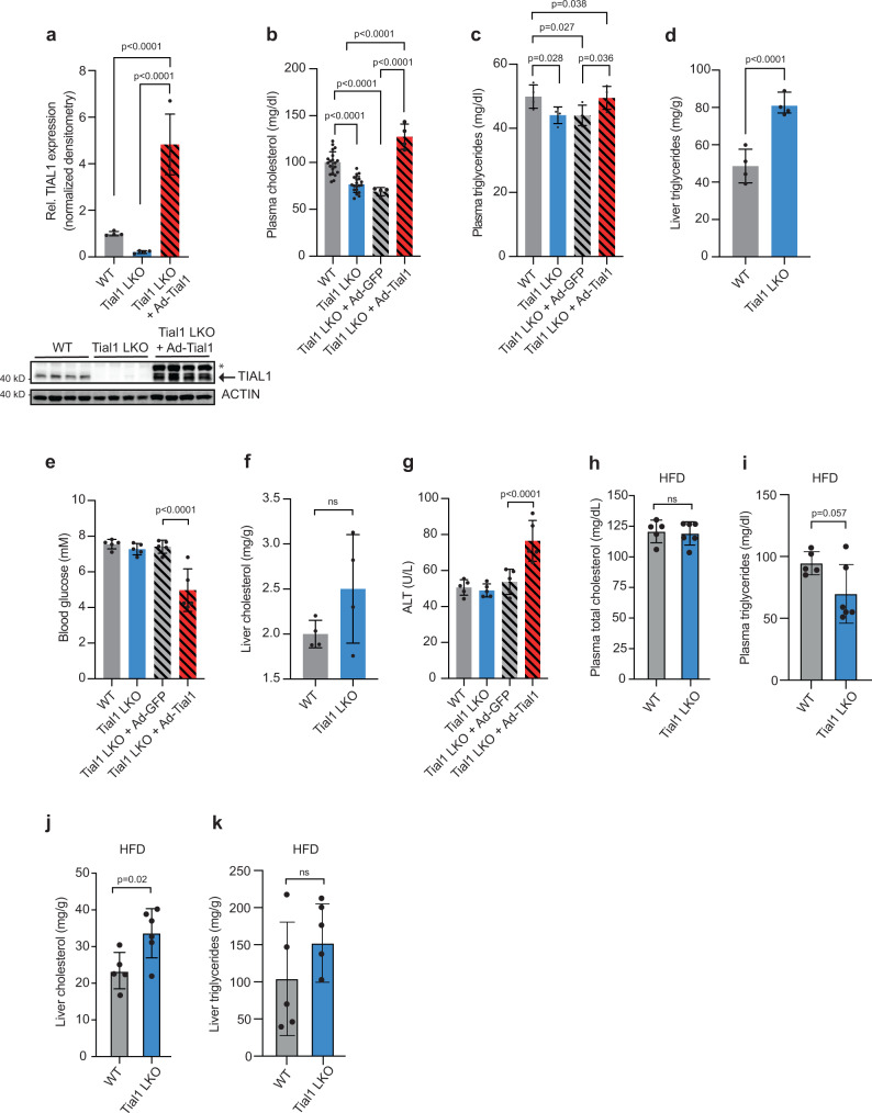
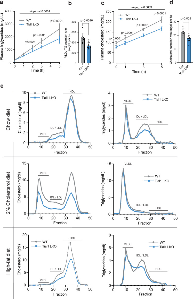
System-wide cross-linking and immunoprecipitation (CLIP) approaches have unveiled regulatory mechanisms of RNA-binding proteins (RBPs) mainly in cultured cells due to limitations in the cross-linking efficiency of tissues. Here, we describe viP-CLIP (in vivo PAR-CLIP), a method capable of identifying RBP targets in mammalian tissues, thereby facilitating the functional analysis of RBP-regulatory networks in vivo. We applied viP-CLIP to mouse livers and identified Insig2 and ApoB as prominent TIAL1 target transcripts, indicating an important role of TIAL1 in cholesterol synthesis and secretion. The functional relevance of these targets was confirmed by showing that TIAL1 influences their translation in hepatocytes. Mutant Tial1 mice exhibit altered cholesterol synthesis, APOB secretion and plasma cholesterol levels. Our results demonstrate that viP-CLIP can identify physiologically relevant RBP targets by finding a factor implicated in the negative feedback regulation of cholesterol biosynthesis.


Conflict of interest statement

The authors declare no competing interests.

Figures

Fig. 1
Fig. 1. A method for determination of RBP-RNA interaction in vivo in mice.
a Schematic representation of in vivo PAR-CLIP (viP-CLIP) applied in mice. Mice are injected 6 times with 4SU over the time course of 12.5 h and sacrificed 15 h after the first dose. Intact organs are flash-frozen, grinded, and cross-linked with UV light (365 nm) under liquid nitrogen cooling. After cell lysis of the tissue powder, the RNA-RBP complexes are immunoprecipitated in presence of RNase, radiolabeled and the recovered RNA in the size of 19–35 nt is ligated to sequencing adapter, and used for cDNA library preparation and sequencing. b 4SU incorporation rates in tissues (n = 3) after injection regime displayed in (a). HEK293 (n = 3) and primary hepatocytes (n = 3) were cultured in the presence of 100 mM 4SU for 16 h and subjected together with the tissue samples to HPLC analysis of 4SU incorporation rates. Values are mean +/– S.D. c Phosphorimage of urea-PAGE transferred to a nitrocellulose membrane that resolved 32P-labeled RNA-TIAL1 complexes after immunoprecipitation of TIAL1 in several tissues. d Immunoblot showing tissue distribution of TIAL1 in mouse organs. e Autoradiograph of recovered RNAs as displayed in (c), after proteinase K treatment and 15% urea gel electrophoresis. 5’ radiolabeled synthetic RNAs of 19 and 35 nt length served as size markers. c, d, e Representative images of three independent replicates. f Graphic presentation of viP-CLIP cDNA library composition by RNA categories from recovered RNA from (e). Source data are provided as a Source Data file.
Fig. 2
Fig. 2. TIAL1 crosslinks and binding to target mRNAs.
a Crosslinked Tial1 liver viP-CLIP reads harboring characteristic T-to-C conversions primarily map to the precursor mRNA and mRNA categories. b PARalyzer-based binding sites distribution in various RNA-species primarily indicates intronic and 3’UTR binding. c Normalized density distribution of Tial1 liver viP-CLIP binding sites (red line) over 3’UTRs compared to a randomized background (gray line). Tial1 binding accumulates close to the poly adenylation and cleave sites. Sequence logo for the RNA recognition element of Tial1 top 1000 3’UTR sequence read clusters is indicated in the graph. d Normalized density distribution of TIAL1 liver viP-CLIP binding sites (red line) over introns compared to a randomized background (gray line). An enrichment of TIAL1 binding at 5′ and 3′ splice sites is observed. Sequence logo for the RNA recognition element of Tial1 top 1000 intronic sequence read clusters is indicated in the graph. e Venn diagram with a target overlap of the two liver viP-CLIPs for the endogenous TIAL1 and Ad-Tial1 (n = 8 and n = 5 mice). f Venn diagram with an overlap of binding sites in 3’UTR for both replicates (n = 8 and n = 5 mice). g Scatterplot of normalized crosslinked read counts for overlapping targets of both replicates. Spearman correlation and statistics are depicted. h Scatterplot of T-to-C counts in crosslinked read per gene versus the expression value in FPKM. Spearman correlation and statistics are depicted. i Liver Tial1 viP-CLIP targets identified in the study. j Representative images of TIAL1 stainings in primary human hepatocytes treated with DMSO (vehicle control), AcLDL (120 µM), or Simvastatin (5 µM) for 24 h (n = 3). k Comparisons of cells treated for 46 h with statin (left) and 24 h with statin and then washed and cultured for additional 22 h without statin (n = 3). l TIAL1 immunoblot of the samples in (j) after nucleocytoplasmic fractionation (n = 2). Source data are provided as a Source Data file.
Fig. 3
Fig. 3. Genetic gain- and loss of function mutations in Tial1 affect cholesterol metabolism.
a Relative protein expression of TIAL1 in livers of wildtype (WT), Tial1 LKO and Ad-Tial1 injected Tial1 LKO animals (n = 4 mice per group). Values are relative densitometric readouts normalized to ACTIN. b Plasma total cholesterol (n = 20 mice for WT, n = 19 mice for Tial1 LKO), c plasma triglycerides, d liver triglycerides, e random blood glucose levels, f liver cholesterol, g alanine transaminase (ALT) levels of indicated groups fed a chow diet (n = 4 mice per group in a, b, c, d, f; n = 5 mice per group in e, g). Measurements of high fat diet (HFD) mice of indicated genotype of plasma cholesterol (h), plasma triglycerides (i), liver cholesterol (j) and liver triglycerides (k). Tial1LKO, and Ad-Tial1 and Ad-GFP injected Tial1 LKO animals (n = 5 per group). Data are presented as mean values ± SDs. Statistical significance was evaluated by two-tailed Student’s t-test (in d, f, hk) or two-tailed ANOVA with Holm-Šídâk post hoc analysis (in a, b, c, e, g). Source data are provided as a Source Data file.
Fig. 4
Fig. 4. Hepatic ablation of Tial1 reduces cholesterol biosynthesis through modulating Insig2 translation.
a TIAL1 liver viP-CLIP reads and cluster for Insig2 3’UTR region, aligned to UCSC genome and conservation tracks. Section of strong binding marked in red boxes. b Western blot analysis of TIAL and proteins involved in the INSIG/SREBP pathway in livers of wildtype (WT) and Tial1 LKO mice. c Densitometric analysis of the immunoblots in (b) (n = 3). Additional samples (n = 4) to improve statistical power for HMGCR and INSIG2 are shown in Supplementary Fig. 4a. d qRT-PCR analysis of transcripts involved in Srebp pathway, cholesterol synthesis and uptake, and fatty acid synthesis (n = 3). e Western blot analysis of TIAL1 and proteins involved in the INSIG/SREBP pathway in the livers of mice injected with Ad-GFP or Ad-Tial1 (n = 5). f Densitometric analysis of the immunoblots in (e) (n = 5). g Relative expression of TIAL1 and INSIG2 in three independent Hepa 1–6 Tial1 knockout cell lines. Densitometric analysis of relative INSIG2 immunoblot signals of WT and Tial1 KO cell lines, normalized to ACTIN, is shown on the right. h 3’UTR activity of Insig2 mRNA harboring TIAL1 binding sites in Hepa1–6 wildtype (WT) and KO cells. Data are expressed as relative luciferase activity (RLU) in WT cells transfected with psiCheck2 vector containing Insig2 3’UTR, normalized to 1 (n = 5). i 3’UTR activity of Insig2 mRNA harboring TIAL1 binding sites in WT and Tial1 LKO hepatocytes, infected with Ad-GFP or Ad-Tial1. Data are expressed as relative luciferase activity normalized to WT cells transfected with Ad-GFP and psiCheck2 vector containing Insig2 3’UTR, normalized to 1 (n = 5). j 35S counts from metabolic labeling and immunoprecipitation of TIAL1 target INSIG2 and non-target HUR in primary mouse hepatocytes from wildtype (WT) and Tial1 LKO mice (n = 4). k 35S counts from metabolic labeling if HUR and INSIG2 in Tial1LKO hepatocytes that were infected with control Ad-GFP or Ad-Tial1 (n = 4). Data are presented as mean values ± SDs. Statistical significance was evaluated by two-tailed Student’s t-test (in c, d, f, g, h, j, k) or one-way ANOVA with Holm-Šídâk post hoc analysis (in i). Source data are provided as a Source Data file.
Fig. 5
Fig. 5. Hepatic ablation and overexpression of TIAL1 regulates cholesterol metabolism.
a Plasma from mice in (Fig. 3a), was fractionated by FPLC and lipoproteins were quantified through measurements of (a) cholesterol and (b) triglycerides in each fraction (n = 5 per group). c Selected fractions from (a, b) were subjected to western blot analysis of VLDL/IDL/LDL (ApoB48/100) as well as HDL (ApoA1) markers. d, e 35S counts from metabolic labeling and immunoprecipitation of TIAL1 targets and non-targets (controls, HUR, APOE) in primary mouse hepatocytes of WT and Tial1 LKO animals (n = 4 per group) (d) and their culture media (e). 35S counts from metabolic labeling and immunoprecipitation of TIAL1 targets and non-targets in primary mouse hepatocytes (n = 4 per group) (f) and the media (g) of Tial1 LKO animals, infected with Ad-GFP or Ad-Tial1. Data are presented as mean values ± SDs. Statistical significance was evaluated by two-tailed Student’s t-test. Source data are provided as a Source Data file.
Fig. 6
Fig. 6. Hepatic inactivation of Tial1 reduces triglycerides and cholesterol secretion.
VLDL-TG and cholesterol secretion assays in 8-week-old (WT; n = 8, Tial1-LKO; n = 9) mice after 6 h fasting and intravenously injection of 500 mg/kg tyloxapol to block lipases. Plasma was taken at the indicated time-points and triglycerides (TG) (a) and total cholesterol (TC) levels (c) were determined by enzymatic assay. b VLDL-TG and d cholesterol secretion rates were calculated from the slopes of increased TG and TC for each mouse. e Plasma from mice fed a chow diet, 2% cholesterol diet (n = 6) for 3 days ad libitum or a high-fat diet for 8 weeks (n = 6) was fractionated by FPLC, and lipoproteins were quantified through measurements of cholesterol and triglycerides in each fraction. All data are presented as mean values ± S.D. Statistical significance was evaluated by two-tailed Student’s t-test (b, d) or two-way ANOVA with Holm-Šídâk post hoc analysis (a, c, e). Source data are provided as a Source Data file.

Similar articles

Cited by

References

    1. Gebauer F, Schwarzl T, Valcarcel J, Hentze MW. RNA-binding proteins in human genetic disease. Nat. Rev. Genet. 2021;22:185–198. doi: 10.1038/s41576-020-00302-y. - DOI - PubMed
    1. Gerstberger S, Hafner M, Tuschl T. A census of human RNA-binding proteins. Nat. Rev. Genet. 2014;15:829–845. doi: 10.1038/nrg3813. - DOI - PMC - PubMed
    1. Lukong KE, Chang KW, Khandjian EW, Richard S. RNA-binding proteins in human genetic disease. Trends Genet. 2008;24:416–425. doi: 10.1016/j.tig.2008.05.004. - DOI - PubMed
    1. Hafner M, et al. CLIP and complementary methods. Nat. Rev. Methods Prim. 2021;1:1–23.
    1. Garzia A, Meyer C, Morozov P, Sajek M, Tuschl T. Optimization of PAR-CLIP for transcriptome-wide identification of binding sites of RNA-binding proteins. Methods. 2017;118-119:24–40. doi: 10.1016/j.ymeth.2016.10.007. - DOI - PMC - PubMed