. 2023 Jun 9;13(1):9449.
doi: 10.1038/s41598-023-36660-w.

Loss of SFXN1 mitigates lipotoxicity and predicts poor outcome in non-viral hepatocellular carcinoma

Affiliations

Loss of SFXN1 mitigates lipotoxicity and predicts poor outcome in non-viral hepatocellular carcinoma

Kohei Yagi et al. Sci Rep. .

Abstract

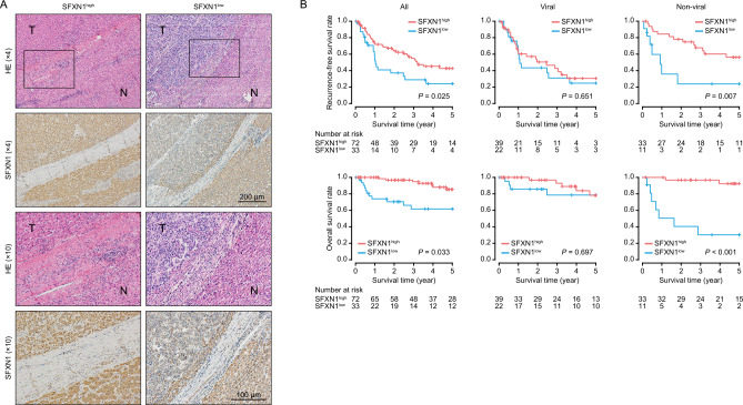
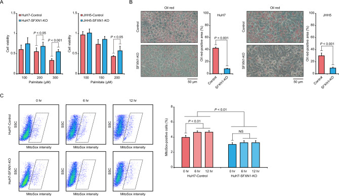
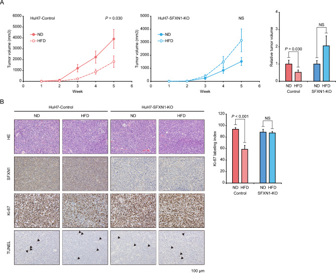
Hepatocellular carcinoma (HCC) imposes a huge global burden, arising from various etiological factors such as hepatitis virus infection and metabolic syndrome. While prophylactic vaccination and antiviral treatment have decreased the incidence of viral HCC, the growing prevalence of metabolic syndrome has led to an increase in non-viral HCC. To identify genes downregulated and specifically associated with unfavorable outcome in non-viral HCC cases, screening analysis was conducted using publically available transcriptome data. Among top 500 genes meeting the criteria, which were involved in lipid metabolism and mitochondrial function, a serine transporter located on inner mitochondrial membrane SFXN1 was highlighted. SFXN1 protein expression was significantly reduced in 33 of 105 HCC tissue samples, and correlated to recurrence-free and overall survival only in non-viral HCC. Human HCC cells with SFXN1 knockout (KO) displayed higher cell viability, lower fat intake and diminished reactive oxygen species (ROS) production in response to palmitate administration. In a subcutaneous transplantation mouse model, high-fat diet feeding attenuated tumorigenic potential in the control cells, but not in the SFXN1-KO cells. In summary, loss of SFXN1 expression suppresses lipid accumulation and ROS generation, preventing toxic effects from fat overload in non-viral HCC, and predicts clinical outcome of non-viral HCC patients.


Conflict of interest statement

The authors declare no competing interests.

Figures

Figure 1
Figure 1
Identification of genes defining patient prognosis in non-viral HCC. (A) Screening analysis of the TCGA data for detecting genes downregulated and specifically associated with poor prognosis in non-viral HCC. Of 8,190 genes satisfying the criteria, 500 genes with the highest difference of P-values between the viral and non-viral HCC groups (ΔlogP = logPviral − logPnon-viral) are designated as “TOP500” genes. Details are described in the Methods and Results sections. (B) DAVID gene ontology analysis of the TOP500 genes. All of the 17 annotations with a FDR < 0.001 are shown. NS: not significant; FDR: false discovery rate.
Figure 2
Figure 2
Evaluation of SFXN1 expression and patient survival in HCC tissues. (A) Representative immunohistochemical images of SFXN1 in HCC specimens. N: adjacent liver tissue; T: tumor tissue; HE: hematoxylin and eosin staining. (B) Kaplan–Meier curves of recurrence-free (upper) and overall (lower) survival in all HCC patients (left; n = 105), viral (middle; n = 61) and non-viral HCC (right; n = 44) groups. P-values are calculated by the log-rank test.
Figure 3
Figure 3
Establishment of the SFXN1-KO HCC cells. (A) mRNA expression analysis of SFXN1 in 26 human HCC cell lines using the Cancer Cell Line Encyclopedia datasets. (B) Protein expression analysis of SFXN1 in immortalized hepatocytes HuSE2 and six human liver cancer cell lines. The SFXN1/GAPDH protein expression ratios are displayed above the blot images. (C) Immunoblot analysis of SFXN1 in the SFXN1-KO HCC cells. (D) Cell proliferation assay of the SFXN1-KO HCC cells. P-values are calculated by Welch’s t-test. Data are the mean ± SD. NS: not significant.
Figure 4
Figure 4
Evaluation of biological effects of SFXN1-KO on HCC cells. (A) Cell viability assay of the SFXN1-KO HCC cells treated with palmitate. P-values are calculated by Welch’s t-test. Data are the mean ± SD. (B) Oil red staining of the SFXN1-KO HCC cells treated with palmitate. The left and right panels show representative images and quantification data of cells stained with oil red, respectively. P-values are calculated by Welch’s t-test. Data are the mean ± SD. (C) Mitochondrial ROS production in response to palmitate administration in the SFXN1-KO HCC cells. The left and right panels show representative flow cytometry plots and quantitative data of the percentage of MitoSox-positive cells, respectively. P-values are calculated by ANOVA with Tukey–Kramer post hoc test. Data are the mean ± SD. SSC: side scatter; NS: not significant.
Figure 5
Figure 5
Evaluation of in vivo effects of SFXN1 attenuation on lipotoxicity. (A) Tumorigenicity assay of the SFXN1-KO HCC cells in immunodeficient mice fed with HFD. The left and right panels show tumor growth curves and relative tumor volumes at five weeks. P-values are calculated by Welch’s t-test. Data are the mean ± SE. (B) Immunohistochemical analysis of tumor tissues derived from the SFXN1-KO HCC cells in immunodeficient mice with HFD. The left and right panels show representative immunohistochemical images and quantitative data of the percentage of Ki-67-positive cells (Ki-67 labeling index), respectively. Arrowheads indicate TUNEL-positive cells. ND: normal diet; HFD: high-fat diet; NS: not significant.

References

    1. Bray F, et al. Global cancer statistics 2018: GLOBOCAN estimates of incidence and mortality worldwide for 36 cancers in 185 countries. CA Cancer J. Clin. 2018;68:394–424. doi: 10.3322/caac.21492. - DOI - PubMed
    1. Fitzmaurice C, et al. Global, regional, and national cancer incidence, mortality, years of life lost, years lived with disability, and disability-adjusted life-years for 29 cancer groups, 1990 to 2017: a systematic analysis for the global burden of disease study. JAMA Oncol. 2019;5:1749–1768. doi: 10.1001/jamaoncol.2019.2996. - DOI - PMC - PubMed
    1. Singal AG, Lampertico P, Nahon P. Epidemiology and surveillance for hepatocellular carcinoma: New trends. J. Hepatol. 2020;72:250–261. doi: 10.1016/j.jhep.2019.08.025. - DOI - PMC - PubMed
    1. Chang MH, et al. Long-term effects of Hepatitis B immunization of infants in preventing liver cancer. Gastroenterology. 2016;151:472–480. doi: 10.1053/j.gastro.2016.05.048. - DOI - PubMed
    1. Yang JD, et al. A global view of hepatocellular carcinoma: Trends, risk, prevention and management. Nat. Rev. Gastroenterol. Hepatol. 2019;16:589–604. doi: 10.1038/s41575-019-0186-y. - DOI - PMC - PubMed

Publication types