

Case report on successful treatment for brain abscess in a Japanese monkey (Macaca fuscata)
- PMID: 37296447
- PMCID: PMC10251556
- DOI: 10.1186/s42826-023-00165-4
Case report on successful treatment for brain abscess in a Japanese monkey (Macaca fuscata)
Retraction in
-
Retraction Note: Case report on successful treatment for brain abscess in a Japanese monkey (Macaca fuscata).Lab Anim Res. 2026 Jan 29;42(1):4. doi: 10.1186/s42826-026-00266-w. Lab Anim Res. 2026. PMID: 41612457 Free PMC article. No abstract available.
Abstract
Background: A brain abscess in human beings is a focal infection of the central nervous system frequently characterized by areas of localized cerebritis and central necrosis surrounded by a well vascularized capsule. A brain abscess, although sporadically reported, is relatively rare disease in domestic animals (horses, cattle, goats and alpacas), companion animals (dogs and cats) and laboratory nonhuman primates. Brain abscesses are life threatening disease that needs early and aggressive veterinary therapy.
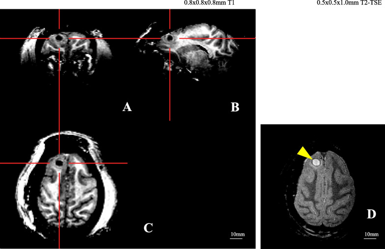
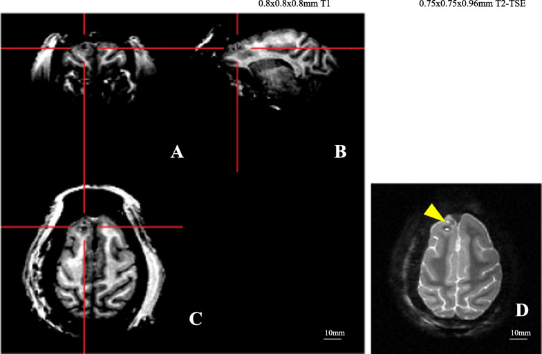
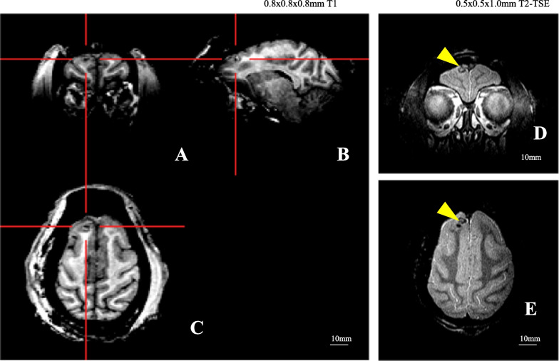
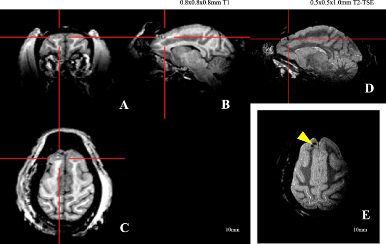
Case presentation: The purpose of this study on a brain abscess in a Japanese monkey was to report the investigational and therapeutic processes including clinical observations, hematological and serum biochemical profiles, and magnetic resonance imaging (MRI) features, probiotic and antibiotic therapy. In clinical observation, the monkey presented with slowly progressive gentle and depressed behavioral change. Hematological findings showed that slightly declined platelet counts gradually increased in the course of the treatment. Serum biochemical profiles revealed initial markedly elevated. A series of chemotherapy provide prominent relief from the influence of the brain abscess. MRI images illustrated that a brain abscess was located in the right frontal lobe and the mass was delineated by a thick rim, indicating the capsule formation stage. The lesion chronologically decreased in size over the course of treatment. Until 11 weeks after treatment of the brain abscess, the size of brain abscess continued to reduce, leaving an organized lesion trace. To the best of my knowledge, this is the first report on successful treatment for a brain abscess in a Japanese monkey (Macaca fuscata).
Conclusions: Medical management of simian brain abscesses is possible based on the controlled and resolving nature of the lesions as determined by MRI and completion of a of chemical antibiotic treatment presented in this study.
Keywords: Antibiotics therapy; Brain abscess; Chemotherapy; Hematology; Japanese monkey (Macaca fuscata); Magnetic resonance imaging; Serum biochemistry.
© 2023. The Author(s).
Conflict of interest statement
The author of this manuscript has no competing interests.
Figures
References
-
- Ruiz-Barrera MA, Santamaria-Rodriguez AF, Zorro OF. Brain abscess: a narrative review. Neurol Perspect. 2022;2(3):160–167. doi: 10.1016/j.neurop.2022.01.010. - DOI
Publication types
LinkOut - more resources
Full Text Sources
Miscellaneous
