. 2023 Jun 9;20(1):139.
doi: 10.1186/s12974-023-02822-w.

Kinesin-5 inhibition improves neural regeneration in experimental autoimmune neuritis

Affiliations

Kinesin-5 inhibition improves neural regeneration in experimental autoimmune neuritis

Felix Kohle et al. J Neuroinflammation. .

Abstract

Background: Autoimmune neuropathies can result in long-term disability and incomplete recovery, despite adequate first-line therapy. Kinesin-5 inhibition was shown to accelerate neurite outgrowth in different preclinical studies. Here, we evaluated the potential neuro-regenerative effects of the small molecule kinesin-5 inhibitor monastrol in a rodent model of acute autoimmune neuropathies, experimental autoimmune neuritis.

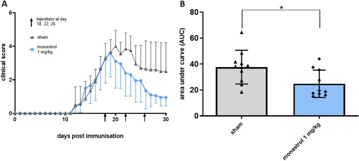
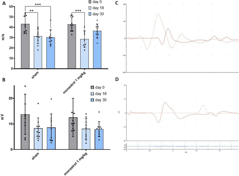
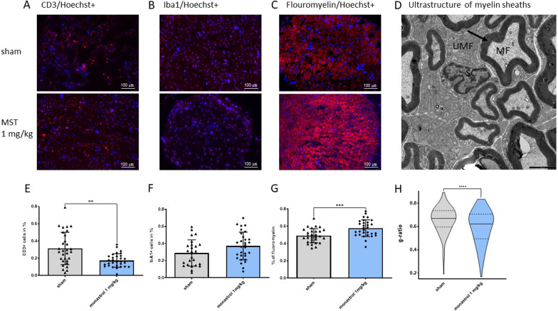
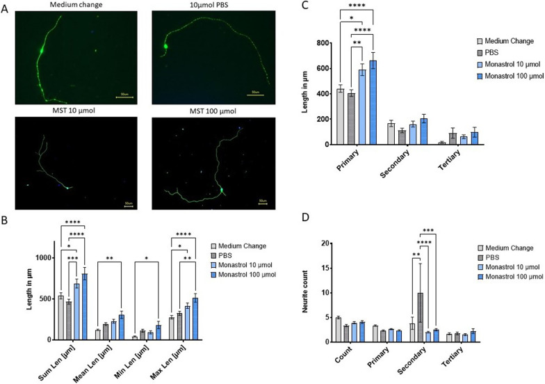
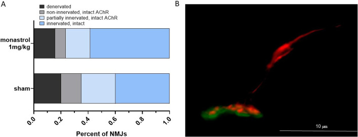
Methods: Experimental autoimmune neuritis was induced in Lewis rats with the neurogenic P2-peptide. At the beginning of the recovery phase at day 18, the animals were treated with 1 mg/kg monastrol or sham and observed until day 30 post-immunisation. Electrophysiological and histological analysis for markers of inflammation and remyelination of the sciatic nerve were performed. Neuromuscular junctions of the tibialis anterior muscles were analysed for reinnervation. We further treated human induced pluripotent stem cells-derived secondary motor neurons with monastrol in different concentrations and performed a neurite outgrowth assay.

Results: Treatment with monastrol enhanced functional and histological recovery in experimental autoimmune neuritis. Motor nerve conduction velocity at day 30 in the treated animals was comparable to pre-neuritis values. Monastrol-treated animals showed partially reinnervated or intact neuromuscular junctions. A significant and dose-dependent accelerated neurite outgrowth was observed after kinesin-5 inhibition as a possible mode of action.

Conclusion: Pharmacological kinesin-5 inhibition improves the functional outcome in experimental autoimmune neuritis through accelerated motor neurite outgrowth and histological recovery. This approach could be of interest to improve the outcome of autoimmune neuropathy patients.

Keywords: Autoimmune neuropathy; Eg5; Experimental autoimmune neuritis; Guillain–Barré syndrome; Monastrol; Neuroregeneration.


Conflict of interest statement

GRF received royalties from Springer, Thieme and Hogrefe. He declared speaker honoraria from Bayer, Desitin, Ergo DKV, Forum für medizinische Fortbildung (FomF) GmbH, GSK, Medica Academy Messe Düsseldorf, Medicbrain Healthcare, Novartis, Pfizer, and Sportärztebund NRW. HCL received honoraria for speaking and advisory board engagement or academic research support by Akcea, Alnylam, Biogen, Celgene, CSL Behring, Grifols, Gruenenthal, LFB Pharma, Takeda and UCB. The other authors declare no conflict of interests.

Figures

Fig. 1
Fig. 1
Treatment with 1 mg/kg monastrol enhances functional recovery after experimental autoimmune neuritis (EAN). EAN score from day 0 to day 30 p.i. is depicted in A. Overall, n = 10 animals were treated in each group with either 1 × PBS as control or 1 mg/kg monastrol i.p. injections at days 18, 22 and 26. Monastrol improves the EAN recovery course. Kinesin-5 inhibition reduces the area under the curve (AUC), shown in B. One-sample t-test was performed to row stats of data analysis, with Mann–Whitney test of AUC values. The experiment was performed three times. The SD is depicted in every picture
Fig. 2
Fig. 2
Monastrol accelerates motor nerve conduction velocity (mNCV) recovery. A Depicts the impact of experimental autoimmune neuritis (EAN) on mNCV at day 0, 18 and 30. The measurements of the compound action muscle potential (cMAP) of the dorsal foot are displayed in B. Both groups showed no significant reduction of cMAP (control group: p-value of comparing days 0 and 18: 0.08, days 0 and 30: 0.11; respective p-values of the monastrol group: 0.21 and 0.17; multiple 2-way ANOVA with Tukey’s multiple comparisons). C and D Show exemplary measurements of mNCV after proximal and distal stimulation at day 0 in a healthy rat in C and at day 18 with a prolonged mNCV in D. Multiple 2-way ANOVA test with Tukey’s multiple comparisons was performed. Overall, n = 10 rats were used. The experiment was repeated three times
Fig. 3
Fig. 3
Red blood cell and white blood cell count ratio (RBC/WBC) count at day 30. A Shows the RBC/WBC ratio of the control and the monastrol 1 mg/kg group (n = 4 animals per group). B Shows a standard May–Grünwald–Giemsa staining of a rat treated with monastrol. The red blood cells without a nucleus can be easily identified. Larger white blood cells are marked purple. An unpaired two-tailed t-test was performed. The standard deviation is depicted in A
Fig. 4
Fig. 4
Immunostaining for the cellular inflammation markers CD3+/Iba1+, semi-quantitative myelin-staining and ultrastructural analysis of myelinated fibres. A and E depict the infiltration of CD3+ cells and B and F for Iba1+ cells in the sciatic nerves of the control and monastrol (MST) group. Fluoromyelin staining is shown in C and G. For each performed immunostaining, we analysed n = 25–30 of 10 nerves per group. In D, transmission electron microscopy of the sciatic nerves is shown. Exemplary, a myelinated fibre was marked with MF; unmyelinated fibres with UMF. In the middle of the image, a Schwann cell is depicted (SC). The black arrow indicates the myelin sheath; the scale is shown in black (2500 nm). Overall, 7–10 images of each nerve’s proximal and distal parts were randomly taken in each group (n = 4 nerves per group, resulting in 264 myelin sheaths for control and 186 fibres for MST). Myelinated fibres of MST-treated rats had a significantly lower g-ratio (axon diameter/ fibre diameter) (H, depicted as a truncated violin plot with the median and quartiles). For statistical analysis, an unpaired two-tailed t-test was performed. The standard deviation is depicted in E, F and G
Fig. 5
Fig. 5
Assessment of the outgrowth of human induced pluripotent stem cells-derived secondary motor neurons. Exemplary immune-histochemical staining of the treatment groups is shown in A. MAP2+ staining allows morphological analysis of neurites, and Hoechst was used for cell body staining. In B, the neurite’s overall, mean, minimal and maximal length are shown. Overall neurites length significantly increased compared to controls (medium change: 537.16 ± 37.17 SEM; PBS: 466.47 ± 29.75 SEM) with monastrol (MST) treatment (monastrol 10 µmol: 687.36 ± 53.87 SEM; monastrol 100 µmol: 808.34 ± 75.22 SEM), mainly due to a length increase of the primary neurites as depicted in C (medium change: 441.63 ± 29.18 SEM; PBS: 405.54 ± 26.22 SEM; monastrol 10 µmol: 590.36 ± 46.33 SEM; monastrol 100 µmol: 662.84 ± 63.37 SEM). D Shows the overall neurite count (mean count for medium change: 4.96 ± 0.28 SEM; PBS: 3.33 ± 0.22; monastrol 10 µmol 3.9 ± 0.22 SEM and monastrol 100 µmol 4.05 ± 0.29). Overall, between 100 and 103 neurons were analysed per group. For statistical analysis, multiple 2-way ANOVA with Tukey’s multiple comparisons, was used. SEM is displayed in BD
Fig. 6
Fig. 6
Innervation status of the neuromuscular junctions (NMJs) at the end of the recovery phase. Immunohistochemical analysis via Tuj1 and synaptophysin displayed the pre-synaptic formation, while the post-synapsis was α-bungarotoxin positive. The shift to (partially) reinnervated NMJs is shown in A. In B, an innervated NMJ of the monastrol group is depicted. The post- and pre-synaptic structures are nearly overlapping. Overall, 89–103 NMJs were analysed per group (n = 4 animals per group)

Similar articles

Cited by

References

    1. Lehmann HC, Wunderlich G, Fink GR, Sommer C. Diagnosis of peripheral neuropathy. Neurol Res Pract. 2020;2:20. doi: 10.1186/s42466-020-00064-2. - DOI - PMC - PubMed
    1. Shahrizaila N, Lehmann HC, Kuwabara S. Guillain–Barré syndrome. Lancet. 2021;397:1214–1228. doi: 10.1016/S0140-6736(21)00517-1. - DOI - PubMed
    1. Forsberg A, de Pedro-Cuesta J, Widén Holmqvist L. Use of healthcare, patient satisfaction and burden of care in Guillain-Barre syndrome. J Rehabil Med. 2006;38:230–236. doi: 10.1080/16501970600582997. - DOI - PubMed
    1. Lehmann HC, Meyer Horste G, Kieseier BC, Hartung HP. Pathogenesis and treatment of immune-mediated neuropathies. Ther Adv Neurol Disord. 2009;2:261–281. doi: 10.1177/1756285609104792. - DOI - PMC - PubMed
    1. Gold R, Hartung HP, Toyka KV. Animal models for autoimmune demyelinating disorders of the nervous system. Mol Med Today. 2000;6:88–91. doi: 10.1016/S1357-4310(99)01639-1. - DOI - PubMed

LinkOut - more resources