. 2023 May 23;12(11):1455.
doi: 10.3390/cells12111455.

Whole-Exome Sequencing Identifies Homozygote Nonsense Variants in LMOD2 Gene Causing Infantile Dilated Cardiomyopathy

Affiliations

Whole-Exome Sequencing Identifies Homozygote Nonsense Variants in LMOD2 Gene Causing Infantile Dilated Cardiomyopathy

Reiri Sono et al. Cells. .

Abstract

As an essential component of the sarcomere, actin thin filament stems from the Z-disk extend toward the middle of the sarcomere and overlaps with myosin thick filaments. Elongation of the cardiac thin filament is essential for normal sarcomere maturation and heart function. This process is regulated by the actin-binding proteins Leiomodins (LMODs), among which LMOD2 has recently been identified as a key regulator of thin filament elongation to reach a mature length. Few reports have implicated homozygous loss of function variants of LMOD2 in neonatal dilated cardiomyopathy (DCM) associated with thin filament shortening. We present the fifth case of DCM due to biallelic variants in the LMOD2 gene and the second case with the c.1193G>A (p.W398*) nonsense variant identified by whole-exome sequencing. The proband is a 4-month male infant of Hispanic descent with advanced heart failure. Consistent with previous reports, a myocardial biopsy exhibited remarkably short thin filaments. However, compared to other cases of identical or similar biallelic variants, the patient presented here has an unusually late onset of cardiomyopathy during infancy. Herein, we present the phenotypic and histological features of this variant, confirm the pathogenic impact on protein expression and sarcomere structure, and discuss the current knowledge of LMOD2-related cardiomyopathy.

Keywords: DCM; LMOD2; heart maturation; leiomodins; neonatal cardiomyopathy; sarcomere; thin filament; whole-exome sequencing; whole-genome sequencing.


Conflict of interest statement

The authors declare no conflict of interest. The funders had no role in the design of the study; in the collection, analyses, or interpretation of data; in the writing of the manuscript, or in the decision to publish the results.

Figures

Figure 1
Figure 1
Trends of Variant Discovery Rate in Neonatal and Infantile Dilated Cardiomyopathy (DCM). ClinVar variant discovery by year, filtered with the exact keywords: (A) “neonatal dilated cardiomyopathy”, (B) “infantile dilated cardiomyopathy”. The number of pathogenic or likely pathogenic (P/LP) variants and variants of uncertain significance (VUS) show a large year-to-year variation with an overall accelerating trend.
Figure 2
Figure 2
Clinical Presentation of The Proband. (A) Chest X-ray on admission demonstrating severe cardiomegaly. L: Left. (B) Representative echocardiogram of LV systolic function (M-Mode) at the level of papillary muscles during systole (a) and diastole (b), demonstrating global left ventricular hypokinesis with estimated EF of 35% and fractional shortening (FS) of 16.5% and significant decrease in left ventricle wall thickness. Please see the accompanied Supplementary Videos included in Supplementary Materials. (C,D) Representative electrocardiogram recording at the baseline (C) on admission and during ventricular tachycardia episode (D) on day 11 of admission.
Figure 3
Figure 3
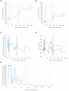
Summary of Cardiac Function Parameters Throughout the Hospital Course. (A) Representing graph summary of percentile left ventricle ejection fraction (LVEF%) measured by the unidimensional mode (M-mode). (B) Representing graph summary of percentile left ventricle fractional shortening (LVFS%) measured by the unidimensional mode (M-mode). (C) Representing graph summary of heart rate. (D) Representing graph summary of T I QT nterval (circles) and corrected QT (QTc, crosses), with line break representing the arrhythmia event where these parameters were not measurable. (E) Representative graph summary of serum brain-natriuretic peptide (BNP); plateau at 5000 pg/mL represents the upper limit of quantification. For all panels, the vertical lines, from left to right, are dates of placement of ECMO (day 13, fine broken line), LVAD (day 15, coarse broken line), and cardiac transplant (day 75, solid line). As of 11 months of age, the patient has recovered from OHT and sustained gaining weight and height appropriately with occasional mild infections. Echocardiogram has demonstrated stable graft function.
Figure 4
Figure 4
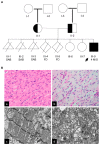
Family Pedigree and Histopathology. (A) Three-generation family pedigree: III-9, Index case; II-1, healthy mother carries the c.1193G>A (p. Trp398*) variant; and II-2, healthy father carries the c.1193G>A (p.Trp389*) variant. III-7 and III-8, older healthy sisters that have not been tested. No other family history of heart disease. III-9, male proband (black arrow), presented at 4 months of age with cardiomegaly and profound heart failure. SAB, spontaneous abortion; FD, Fetal Demise. (B) Histology of myocardial tissue obtained during left ventricle assist devise implantation (Pre-LVAD). (a) Mild cellular vacuolization on light microscopy (H&E stain, 400× magnification); (b) Periodic Acid Schiff stain showed mild increase in glycogen (PAS stain, 400× magnification); (c) electron microscopy representative imaging (×10,000) depicts disorganized Z-bands, separation of sarcomeres by edema, and rare cytosolic glycogen; no significant lysosome-bound glycogen is noted; (d) electron microscopy representative imaging (×50,000) of left ventricular biopsy depicts focal increase in mitochondria with nonspecific mitochondrial swelling and pleoconia noted, with no significant cristae abnormality or inclusions.
Figure 5
Figure 5
Homozygous Loss of Function Variant of LMOD2 Revealed by Trio (Proband/Parents) Whole Exome Sequencing (WES). (A) Table summary depicts rare heterozygous variants of uncertain significance in PKP2, SCN5A, and SLC37A4 genes and homozygous loss of function variant in LMOD2 detected by WES. (B) Integrated genomic viewer (IGV) windows depict homozygous c.1193G>A variant in LMOD2 in the proband genome, while both parents are heterozygous carriers. Green represents the adenine variant; brown represents guanine base (wildtype). (C) * Schematic representation of Lmod2 protein with known structural motifs (upper, adopted from Chen et al. 2015 [22]) and LMOD2 gene (lower). Black arrows indicate exons affected by previously reported deleterious variants. p.W398* indicates the position of the substitution in the present proband caused by the c.1193G>A variant. Please see accompanied Supplementary Figure S1 in Supplementary Material.
Figure 6
Figure 6
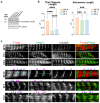
Pathologic Impact of LMOD2 c.1193G>A (p.W398*) Variant. (A) Immunoblot of Lmod2 protein in proband explant left ventricle (LV) and right ventricle (RV) affected with homozygous LMOD2 c.1193G>A (p.W398*) and in LV and RV in an age-matched control. Gapdh was used as a loading control. (B) Thin filament lengths and sarcomere lengths in LV measured by Tmod1 staining of the proband (Pt.2), a 14-month-old non-failing control (NF14), and the first case reported with the homozygous LMOD2 c.1193G>A (p.W398*) variant (Pt.1). Measurements were obtained using the DDecon ImageJ plugin. Values are mean ± SD, and n = 44, 22, and 37 measurements (NF14, Pt.1, and Pt.2, respectively). *** p < 0.001 and **** p < 0.0001, one-way ANOVA. ns: non-significant (C) Representative immunofluorescence images of LV from NF14, Pt.1, and Pt.2. Fluorescently labeled phalloidin stains F-actin (red), Tmod1 staining marks pointed ends (green), and α-actinin marks Z-discs (Z). Magenta arrows mark thin filament pointed ends, and cyan lines show examples of thin filament arrays used for measurements. Scale bar: 2 µm.

References

    1. Mestroni L., Brun F., Spezzacatene A., Sinagra G., Taylor M.R. Genetic causes of dilated Cardiomyopathy. Prog. Pediatr. Cardiol. 2014;37:13–18. doi: 10.1016/j.ppedcard.2014.10.003. - DOI - PMC - PubMed
    1. Soares P., Rocha G., Pissarra S., Soares H., Flôr-de-Lima F., Costa S., Moura C., Dória S., Guimarães H. Neonatal Dilated Cardiomyopathy. Rev. Port. Cardiol. 2017;36:201–214. doi: 10.1016/j.repc.2016.10.007. - DOI - PubMed
    1. Copel J.A., Cullen M., Green J.J., Mahoney M.J., Hobbins J.C., Kleinman C.S. The Frequency of Aneuploidy in Prenatally Diagnosed Congenital Heart Disease: An Indication for Fetal Karyotyping. Am. J. Obstet. Gynecol. 1988;158:409–413. doi: 10.1016/0002-9378(88)90166-4. - DOI - PubMed
    1. Pappas C.T., Mayfield R.M., Henderson C., Jamilpour N., Cover C., Hernandez Z., Hutchinson K.R., Chu M., Nam K.-H., Valdez J.M., et al. Knockout of Lmod2 Results in Shorter Thin Filaments Followed by Dilated Cardiomyopathy and Juvenile Lethality. Proc. Natl. Acad. Sci. USA. 2015;112:13573–13578. doi: 10.1073/pnas.1508273112. - DOI - PMC - PubMed
    1. Pappas C.T., Farman G.P., Mayfield R.M., Konhilas J.P., Gregorio C.C. Cardiac-Specific Knockout of Lmod2 Results in a Severe Reduction in Myofilament Force Production and Rapid Cardiac Failure. J. Mol. Cell Cardiol. 2018;122:88–97. doi: 10.1016/j.yjmcc.2018.08.009. - DOI - PMC - PubMed

Publication types