. 2023 May 31;12(11):1522.
doi: 10.3390/cells12111522.

In Situ Identification of Both IL-4 and IL-10 Cytokine-Receptor Interactions during Tissue Regeneration

Affiliations

In Situ Identification of Both IL-4 and IL-10 Cytokine-Receptor Interactions during Tissue Regeneration

Krisztina Nikovics et al. Cells. .

Abstract

Cytokines secreted by individual immune cells regulate tissue regeneration and allow communication between various cell types. Cytokines bind to cognate receptors and trigger the healing process. Determining the orchestration of cytokine interactions with their receptors on their cellular targets is essential to fully understanding the process of inflammation and tissue regeneration. To this end, we have investigated the interactions of Interleukin-4 cytokine (IL-4)/Interleukin-4 cytokine receptor (IL-4R) and Interleukin-10 cytokine (IL-10)/Interleukin-10 cytokine receptor (IL-10R) using in situ Proximity Ligation Assays in a regenerative model of skin, muscle and lung tissues in the mini-pig. The pattern of protein-protein interactions was distinct for the two cytokines. IL-4 bound predominantly to receptors on macrophages and endothelial cells around the blood vessels while the target cells of IL-10 were mainly receptors on muscle cells. Our results show that in situ studies of cytokine-receptor interactions can unravel the fine details of the mechanism of action of cytokines.

Keywords: IL-10; IL-4; interaction; proximity ligation assay; receptor; regeneration.


Conflict of interest statement

The authors declare no conflict of interest.

Figures
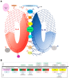
Figure 1
Figure 1
(A) Interleukin-4 (IL-4) and interleukin-10 (IL-10) are cytokines secreted by different cells that play an important role in the regulation of immune responses. They have specific receptors through which they exert their effects. Below, we briefly review the cells expressing IL-4 (red asterisks) and IL-10 (blue asterisks) and their receptors IL-4R (red receptor) and IL-10R (blue receptor). There are four types of cells (granulocytes, T cells, mast cells, and macrophages) that produce both cytokines and their receptors. Four cell types (DC cells, NK cells, B cells, and monocytes) secrete only IL-10, but both receptors can be detected on the surface. ILC2 cells express IL-4. The IL-4 receptor is located on the surface of fibroblasts and muscle cells, while both receptors are present on the surface of endothelial cells. In addition, there are cells (marked with a question mark) that produce these cytokines and their receptors, but their identity has not yet been determined. (B) Strategy to develop and validate a protein–protein interaction using the PLA method. First, macrophage detection was performed on the tissue of interest; second, actors were identified by immunofluorescence before performing the PLA method to detect protein–protein interaction. Blue boxes represent figures that contain both wounded and healthy tissue. The brown boxes correspond to wounded tissue and the black boxes to healthy tissue. Green boxes indicate negative controls.
Figure 2
Figure 2
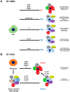
Activation and classification of macrophages: in vitro and in vivo macrophage polarization depending on the activation pathway. (A) In vitro, M0 macrophages can be transformed into different phenotypes depending on the activation pathway. This polarization is influenced by different molecules (including several cytokines). IL-4/IL-13 induces polarization of M2a, IL-1R agonists/Toll-like receptor induces polarization of M2b, IL-10 induces polarization of M2c, IL-6 induces polarization of M2d macrophages, in contrast to IFN-γ, TNF, and Lipopolysaccharide (LPS), which induce polarization of M1 macrophages. Understanding the details of polarization is essential as different subtypes have different functions. (B) In contrast, the polarization of macrophages in vivo is relatively poorly understood. Further studies are needed to determine which molecules are important in the polarization of the macrophage types involved. M0: non-activated macrophage.
Figure 3
Figure 3
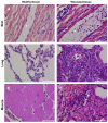
Macroscopic morphology of tissues. Histologic HPS staining was performed on (A,B) skin (C,D) lung, and (E,F) muscle sections in (A,C,E) healthy tissue and (B,D,F) wounded tissue. Scale bar = 50 µm. White arrow: granulocyte; yellow arrow: macrophage; star: lumen of the blood vessel.
Figure 4
Figure 4
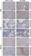
Identification of tissue-resident and M2-like macrophages in situ. (B,D,F,H,J,L) Immunolabeling with CD206 of the (AD) skin of the (EH) lung and the (IL) muscle sections. (A,B,E,F,I,J) Healthy tissue. (C,D,G,H,K,L) Wounded tissue. (A,C,E,G,I,K) Negative controls of the immunolabeling. (B,D,F,H,J,L) Anti-CD206 (brown color) labeling the tissue-resident and M2-like macrophages. Nuclear staining with hematoxylin (blue color). Scale bar = 100 µm. White arrow: Tissue-resident macrophage; red arrow: M2-like macrophage.
Figure 5
Figure 5
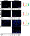
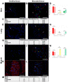
Identification of IL-4R+ and CD206+ cells in situ. (A,B) Immunolabeling of both IL-4R and CD206 in the skin, (E,F) the muscle, and (IK) the lung sections. (A,E,I) Healthy tissue. (B,F,J,K) Wounded tissue. (C,D) Quantitative analysis of both IL-4R+ and CD206+ cells in the skin; (G,H) the muscle and (L,M) the lung. Quantitative analysis based on random examination of 3 sets of 1000 cells per condition. Anti-IL-4R (Alexa488, green fluorescence) and Anti-CD206 (Alexa568, red fluorescence) label the tissue-resident and M2-like macrophages. Nuclear staining with DAPI (blue fluorescence). Scale bar = 50 µm. Yellow arrow: endothelial cell; white arrow: macrophage; and red arrow: IL-4R + cell. ** p < 0.01, * p < 0.5, ns = not significant, compared to healthy tissue.
Figure 6
Figure 6
Identification of IL-10R+ and CD206+ cells in situ. (A,B) Immunolabeling of both IL-10R and CD206 in the skin, (E,F) the lung, and (IL) the muscle sections. (L) identical to the K image, including the autofluorescence of muscle cells. (A,E,I) Healthy tissue. (B,F,J,K,L) Wounded tissue. (C,D) Quantitative analysis of the IL-10R+ and CD206+ cells in the skin; (G,H) the lung and (M,N) the muscle. Quantitative analysis based on random examination of 3 sets of 1000 cells per condition. Anti-IL-10R (Alexa488, green fluorescence) and Anti-CD206 (Alexa568, red fluorescence) label the tissue-resident and M2-like macrophages. Nuclear staining with DAPI (blue fluorescence). Scale bar = 50 µm. Yellow arrow: endothelial cell; white arrow: macrophage; and red arrow: IL-10R+ cell. ** p < 0.01, * p < 0.5, ns = not significant, compared to healthy tissue.
Figure 7
Figure 7
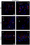
Identification of IL-4R+ and IL-4+cells in situ. (A,B) Immunolabeling of both IL-4R and IL-4 in the wounded skin, (C,D) the lung, and (E,F) the muscle sections. (B,D,F) Expanded view: high magnification image of the area within the red rectangle in A,C, and E, respectively. Anti-IL-4R (Alexa488, green fluorescence) and Anti-IL-4 (Alexa568, red fluorescence). Nuclear staining with DAPI (blue fluorescence). Scale bar = 20 µm. Star: lumen of the blood vessel; white arrow: IL-4+ granulocyte; and yellow arrow: IL-4R+/IL-4+ cell.
Figure 8
Figure 8
Identification of IL-10R+ and IL-10+ cells in situ. (A,B) Immunolabeling of both anti-IL-10R and IL-10 antibodies in the wounded skin, (C,D) the lung, and (E,F) the muscle sections. (B,D) Expanded view: high magnification image of the area within the red rectangle in (A,C), respectively. (F) Identical to the E image, including the autofluorescence of muscle cells. Anti-IL-10R (Alexa488, green fluorescence) and Anti-IL-10 (Alexa568, red fluorescence). Nuclear staining with DAPI (blue fluorescence). Scale bar = 20 µm. Star: lumen of the blood vessel; white arrow: IL-10R+ cell; yellow arrow: IL-10+ cell; and red arrow: granulocyte in the blood vessel.
Figure 9
Figure 9
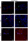
Interaction between IL-4 and IL-4R in situ. (A,B) Proximity Ligation Assay with anti-IL-4 and anti-IL-4R antibodies in the wounded skin, (C,D) the wounded lung, and (E,F) the wounded muscle. (B,F) Expanded view: high magnification image of the area within the red rectangle in A,E. Alexa568, yellow fluorescence, labeling the interaction between IL-4 and IL-4R. Nuclear staining with DAPI (blue fluorescence). Scale bar = 20 µm. Star: lumen of the blood vessel; white arrow: Proximity Ligation Assay IL-4R+/IL-4+ cells; and yellow arrow: granulocyte.
Figure 10
Figure 10
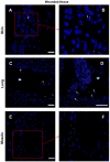
Interaction between IL-10 and IL-10R in situ. (A,B) Proximity Ligation Assay with anti-IL-10R and anti-IL-10 in the wounded skin (C,D) the wounded lung and (E,F) the wounded muscle. (B,D) Expanded view: high magnification image of the area within the red rectangle in (A,C). Alexa568, yellow fluorescence, labeling the interaction between IL-10 and IL-10R. Nuclear staining with DAPI (blue fluorescence). Scale bar = 20 µm. Star: lumen of the blood vessel; white arrow: Proximity Ligation Assay IL-10R/IL-10+ cells; and yellow arrow: muscle fiber.
Figure 11
Figure 11
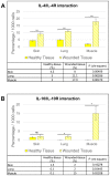
Quantitative analysis of both (A) IL-4/IL-4R and (B) IL-10/IL-10R interactions. Quantitative analysis based on random examination of 3 sets of 1000 cells per condition. ** p < 0.01, * p < 0.5, ns = not significant, compared to healthy tissue.
Figure 12
Figure 12
Summary of our results on the in situ identification of IL-4 and IL-10 cytokine receptor interactions. Endothelial cells showed intense IL-4 cytokine receptor interaction and muscle cells IL-10 cytokine receptor interaction during regeneration. The presence of IL-4 was detected in granulocytes and both receptors in macrophages. Contrary to the literature, the presence of IL-10R in endothelial cells and IL-4R in muscle cells was not detected. In addition, we identified further cells producing cytokines or their receptors (marked with a question mark), which will be characterized in the future. IL-4 is shown in red and IL-10 in blue. Interactions detected are highlighted in yellow.

References

    1. Martinez F.O., Gordon S. The M1 and M2 Paradigm of Macrophage Activation: Time for Reassessment. F1000Prime Rep. 2014;6:13. doi: 10.12703/P6-13. - DOI - PMC - PubMed
    1. Snyder R.J., Lantis J., Kirsner R.S., Shah V., Molyneaux M., Carter M.J. Macrophages: A Review of Their Role in Wound Healing and Their Therapeutic Use. Wound Repair. Regen. 2016;24:613–629. doi: 10.1111/wrr.12444. - DOI - PubMed
    1. Abdelaziz M.H., Abdelwahab S.F., Wan J., Cai W., Huixuan W., Jianjun C., Kumar K.D., Vasudevan A., Sadek A., Su Z., et al. Alternatively Activated Macrophages; a Double-Edged Sword in Allergic Asthma. J. Transl. Med. 2020;18:58. doi: 10.1186/s12967-020-02251-w. - DOI - PMC - PubMed
    1. Xu H.-T., Lee C.-W., Li M.-Y., Wang Y.-F., Yung P.S.-H., Lee O.K.-S. The Shift in Macrophages Polarisation after Tendon Injury: A Systematic Review. J. Orthop. Translat. 2020;21:24–34. doi: 10.1016/j.jot.2019.11.009. - DOI - PMC - PubMed
    1. Arora S., Dev K., Agarwal B., Das P., Syed M.A. Macrophages: Their Role, Activation, and Polarization in Pulmonary Diseases. Immunobiology. 2018;223:383–396. doi: 10.1016/j.imbio.2017.11.001. - DOI - PMC - PubMed

Publication types

LinkOut - more resources