. 2023 Jun 2;12(11):1537.
doi: 10.3390/cells12111537.

Short-Term Exposure to Bisphenol A Does Not Impact Gonadal Cell Steroidogenesis In Vitro

Affiliations

Short-Term Exposure to Bisphenol A Does Not Impact Gonadal Cell Steroidogenesis In Vitro

Neena Roy et al. Cells. .

Abstract

Bisphenol A (BPA) is a ubiquitous, synthetic chemical proven to induce reproductive disorders in both men and women. The available studies investigated the effects of BPA on male and female steroidogenesis following long-term exposure to the compound at relatively high environmental concentrations. However, the impact of short-term exposure to BPA on reproduction is poorly studied. We evaluated if 8 and 24 h exposure to 1 nM and 1 µM BPA perturbs luteinizing hormone/choriogonadotropin (LH/hCG)-mediated signalling in two steroidogenic cell models, i.e., the mouse tumour Leydig cell line mLTC1, and human primary granulosa lutein cells (hGLC). Cell signalling studies were performed using a homogeneous time-resolved fluorescence (HTRF) assay and Western blotting, while gene expression analysis was carried out using real-time PCR. Immunostainings and an immunoassay were used for intracellular protein expression and steroidogenesis analyses, respectively. The presence of BPA leads to no significant changes in gonadotropin-induced cAMP accumulation, alongside phosphorylation of downstream molecules, such as ERK1/2, CREB and p38 MAPK, in both the cell models. BPA did not impact STARD1, CYP11A1 and CYP19A1 gene expression in hGLC, nor Stard1 and Cyp17a1 expression in mLTC1 treated with LH/hCG. Additionally, the StAR protein expression was unchanged upon exposure to BPA. Progesterone and oestradiol levels in the culture medium, measured by hGLC, as well as the testosterone and progesterone levels in the culture medium, measured by mLTC1, did not change in the presence of BPA combined with LH/hCG. These data suggest that short-term exposure to environmental concentrations of BPA does not compromise the LH/hCG-induced steroidogenic potential of either human granulosa or mouse Leydig cells.

Keywords: Bisphenol A (BPA); LH; hCG; ovary; steroidogenesis; testis.


Conflict of interest statement

The authors declare no conflict of interest.

Figures

Figure 1
Figure 1
Effect of BPA on cAMP production in vitro: (A) in human granulosa cells (hGLC); (B) in mouse Leydig cells (mLTC1). cAMP was measured using an HTRF assay. Forskolin and cholera toxin served as the positive control in hGLC and mLTC1 cells, respectively. Data are represented as mean ± SEM. CTX = cholera toxin. *, Significantly different vs. “no gonadotropin” alone; # vs. “no gonadotropin” + BPA 1 nM; $ vs. “no gonadotropin” + BPA 1 µM; N, not significantly different vs. LH/hCG treatment in the absence of BPA (Kruskal–Wallis test, p ≥ 0.05).
Figure 2
Figure 2
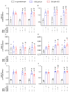
Effect of BPA on activation of pERK, pCREB and p38MAPK: Western blotting analyses in hGLC (A) and in mLTC1 (B) after exposure to BPA without or with hCG or LH. Relative semi-quantification of the activation of pERK (C,D), pCREB (E,F) and p38MAPK (G,H) in hGLC and mLTC1 cells, respectively. Data are represented as mean ± SEM. *, Significantly different vs. “no gonadotropin” alone; # vs. “no gonadotropin” + BPA 1 nM; $ vs. “no gonadotropin” + BPA 1 µM; N, not significantly different vs. LH/hCG treatment in the absence of BPA (Kruskal–Wallis test, p ≥ 0.05).
Figure 3
Figure 3
Effect of BPA on steroidogenic gene expression in hGLC after 8 h (A,C,E) and 24 h (B,D,F) of treatment. Genes: STARD1 (A,B), CYP17A1 (C,D) and CYP19A1 (E,F). Data are represented as mean ± SEM. *, Significantly different vs. “no gonadotropin” alone; # vs. “no gonadotropin” + BPA 1 nM; $ vs. “no gonadotropin” + BPA 1 µM; N, not significantly different vs. LH/hCG treatment in the absence of BPA (Kruskal–Wallis test, p ≥ 0.05).
Figure 4
Figure 4
Effect of BPA on steroidogenic gene expression in mLTC1 after 8 h (A,C) and 24 h (B,D). Genes: Stard1 (A,C) and Cyp17a1 (B,D). Data are represented as mean ± SEM. *, Significantly different vs. “no gonadotropin” alone; # vs. “no gonadotropin” + BPA 1 nM; $ vs. “no gonadotropin” + BPA 1 µM; N, not significantly different vs. LH/hCG treatment in the absence of BPA (Kruskal–Wallis test, p ≥ 0.05).
Figure 5
Figure 5
Immunofluorescence images of the effect of BPA on StAR protein expression in LH/hCG-treated hGLC (AF) and in mLTC1 cells (GL). Cells were treated 24 h with 1 µM of BPA, in the absence or presence of LH/hCG. Images are representative of three independent experiments (magnification 40×. Subfigure size is reduced to 50%).
Figure 6
Figure 6
The effect of BPA on progesterone and oestradiol levels in hGLC cells after 8 h (A,C) and 24 h (B,D). Data are represented as mean ± SEM. *, Significantly different vs. “no gonadotropin” alone; # vs. “no gonadotropin” + BPA 1 nM; $ vs. “no gonadotropin” + BPA 1 µM; N, not significantly different vs. LH/hCG treatment in the absence of BPA (Kruskal–Wallis test, p ≥ 0.05).
Figure 7
Figure 7
The effect of BPA on progesterone and testosterone levels in mLTC1 cells after 8 h (A,C) and 24 h (B,D). Mean ± SEM. *, Significantly different vs. “no gonadotropin” alone; # vs. “no gonadotropin” + BPA 1 nM; $ vs. “no gonadotropin” + BPA 1 µM; N, not significantly different vs. LH/hCG treatment in the absence of BPA (Kruskal–Wallis test, p ≥ 0.05).

References

    1. Peretz J., Vrooman L., Ricke W.A., Hunt P.A., Ehrlich S., Hauser R., Padmanabhan V., Taylor H.S., Swan S.H., Vandevoort C.A., et al. Bisphenol A and reproductive health: Update of experimental and human evidence, 2007–2013. Environ. Health Perspect. 2014;122:775–786. doi: 10.1289/ehp.1307728. - DOI - PMC - PubMed
    1. Rubin B.S. Bisphenol A: An endocrine disruptor with widespread exposure and multiple effects. J. Steroid Biochem. Mol. Biol. 2011;127:27–34. doi: 10.1016/j.jsbmb.2011.05.002. - DOI - PubMed
    1. Staples C.A., Dorn P.B., Klecka G.M., O’Block S.T., Harris L.R. A review of the environmental fate, effects, and exposures of bisphenol A. Chemosphere. 1998;36:2149–2173. doi: 10.1016/S0045-6535(97)10133-3. - DOI - PubMed
    1. Nam S.H., Seo Y.M., Kim M.G. Bisphenol A migration from polycarbonate baby bottle with repeated use. Chemosphere. 2010;79:949–952. doi: 10.1016/j.chemosphere.2010.02.049. - DOI - PubMed
    1. Lim D.S., Kwack S.J., Kim K.B., Kim H.S., Lee B.M. Potential risk of bisphenol a migration from polycarbonate containers after heating, boiling, and microwaving. J. Toxicol. Environ. Health-Part A Curr. Issues. 2009;72:1285–1291. doi: 10.1080/15287390903212329. - DOI - PubMed

Publication types

LinkOut - more resources