

Contemporary Review of Multimodality Imaging of the Prostate Gland
- PMID: 37296712
- PMCID: PMC10252565
- DOI: 10.3390/diagnostics13111860
Contemporary Review of Multimodality Imaging of the Prostate Gland
Abstract
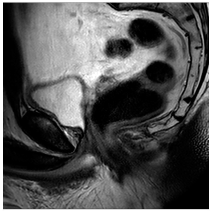
Tissue changes and the enlargement of the prostate, whether benign or malignant, are among the most common groups of diseases that affect men and can have significant impacts on length and quality of life. The prevalence of benign prostatic hyperplasia (BPH) increases significantly with age and affects nearly all men as they grow older. Other than skin cancers, prostate cancer is the most common cancer among men in the United States. Imaging is an essential component in the diagnosis and management of these conditions. Multiple modalities are available for prostate imaging, including several novel imaging modalities that have changed the landscape of prostate imaging in recent years. This review will cover the data relating to commonly used standard-of-care prostate imaging modalities, advances in newer technologies, and newer standards that impact prostate gland imaging.
Keywords: benign prostatic hyperplasia; prostate cancer; prostatic adenocarcinoma.
Conflict of interest statement
The authors declare no conflict of interest relevant to the writing of this manuscript.
Figures


Similar articles
-
Benign prostatic hyperplasia: clinical overview and value of diagnostic imaging.Radiol Clin North Am. 2000 Jan;38(1):31-47. doi: 10.1016/s0033-8389(05)70148-2. Radiol Clin North Am. 2000. PMID: 10664665 Review.
-
Role of mpMRI in Benign Prostatic Hyperplasia Assessment and Treatment.Curr Urol Rep. 2020 Oct 26;21(12):55. doi: 10.1007/s11934-020-01005-x. Curr Urol Rep. 2020. PMID: 33104969 Review.
-
Comprehensive patient evaluation for benign prostatic hyperplasia.Urology. 1998 Apr;51(4A Suppl):13-8. doi: 10.1016/s0090-4295(98)00050-8. Urology. 1998. PMID: 9586591 Review.
-
More advantages in detecting bone and soft tissue metastases from prostate cancer using 18F-PSMA PET/CT.Hell J Nucl Med. 2019 Jan-Apr;22(1):6-9. doi: 10.1967/s002449910952. Epub 2019 Mar 7. Hell J Nucl Med. 2019. PMID: 30843003
-
Surgical Complications in the Management of Benign Prostatic Hyperplasia Treatment.Curr Urol Rep. 2022 May;23(5):83-92. doi: 10.1007/s11934-022-01091-z. Epub 2022 Mar 9. Curr Urol Rep. 2022. PMID: 35262855 Review.
References
-
- Siddiqui M.M., Rais-Bahrami S., Turkbey B., George A.K., Rothwax J., Shakir N., Okoro C., Raskolnikov D., Parnes H.L., Linehan W.M., et al. Comparison of MR/ultrasound fusion-guided biopsy with ultrasound-guided biopsy for the diagnosis of prostate cancer. JAMA. 2015;313:390–397. doi: 10.1001/jama.2014.17942. - DOI - PMC - PubMed
-
- Banker H., Selvarajan S.K. Prostate Imaging, in StatPearls. StatPearls Publishing LLC; Treasure Island, FL, USA: 2023. - PubMed
Publication types
LinkOut - more resources
Full Text Sources
