

Imaging Criteria for the Diagnosis of Progressive Supranuclear Palsy: Supportive or Mandatory?
- PMID: 37296819
- PMCID: PMC10252358
- DOI: 10.3390/diagnostics13111967
Imaging Criteria for the Diagnosis of Progressive Supranuclear Palsy: Supportive or Mandatory?
Abstract
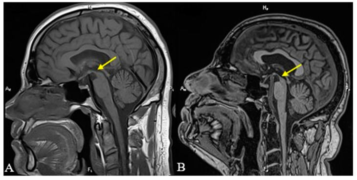
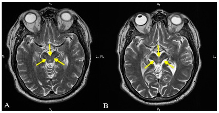
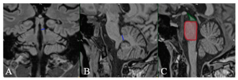
We present the case of a 54-year-old male, without any significant medical history, who insidiously developed speech disturbances and walking difficulties, accompanied by backward falls. The symptoms progressively worsened over time. The patient was initially diagnosed with Parkinson's disease; however, he failed to respond to standard therapy with Levodopa. He came to our attention for worsening postural instability and binocular diplopia. A neurological exam was highly suggestive of a Parkinson-plus disease, most likely progressive supranuclear gaze palsy. Brain MRI was performed and revealed moderate midbrain atrophy with the characteristic "hummingbird" and "Mickey mouse" signs. An increased MR parkinsonism index was also noted. Based on all clinical and paraclinical data, a diagnosis of probable progressive supranuclear palsy was established. We review the main imaging features of this disease and their current role in diagnosis.
Keywords: Magnetic Resonance Parkinsonism Index (MRPI); Mickey mouse sign; PSP syndromes; corticobasal degeneration; hummingbird sign; midbrain atrophy; morning glory sign; multiple system atrophy (MSA); postmortem histopathological examination; progressive supranuclear palsy.
Conflict of interest statement
The authors declare no conflict of interest.
Figures
Similar articles
-
The diagnostic accuracy of the hummingbird and morning glory sign in patients with neurodegenerative parkinsonism.Parkinsonism Relat Disord. 2018 Sep;54:90-94. doi: 10.1016/j.parkreldis.2018.04.005. Epub 2018 Apr 3. Parkinsonism Relat Disord. 2018. PMID: 29643007
-
Morning glory sign: a particular MR finding in progressive supranuclear palsy.Magn Reson Med Sci. 2004 Dec 15;3(3):125-32. doi: 10.2463/mrms.3.125. Magn Reson Med Sci. 2004. PMID: 16093629
-
Progressive supranuclear palsy: A case report and brief review of the literature.Radiol Case Rep. 2023 Nov 2;19(1):250-253. doi: 10.1016/j.radcr.2023.09.012. eCollection 2024 Jan. Radiol Case Rep. 2023. PMID: 38028282 Free PMC article.
-
Progressive Supranuclear Palsy-Parkinsonism Predominant (PSP-P)-A Clinical Challenge at the Boundaries of PSP and Parkinson's Disease (PD).Front Neurol. 2020 Mar 10;11:180. doi: 10.3389/fneur.2020.00180. eCollection 2020. Front Neurol. 2020. PMID: 32218768 Free PMC article. Review.
-
Clinical features differentiating patients with postmortem confirmed progressive supranuclear palsy and corticobasal degeneration.J Neurol. 1999 Sep;246 Suppl 2:II1-5. doi: 10.1007/BF03161075. J Neurol. 1999. PMID: 10525996 Review.
Cited by
-
Automated brain segmentation and volumetry in dementia diagnostics: a narrative review with emphasis on FreeSurfer.Front Aging Neurosci. 2024 Sep 3;16:1459652. doi: 10.3389/fnagi.2024.1459652. eCollection 2024. Front Aging Neurosci. 2024. PMID: 39291276 Free PMC article. Review.
References
-
- Allan H.R., Martin A.S., Joshua P.K., Sashank P., editors. Adams and Victor’s Principles of Neurology. McGraw-Hill Educational; New York, NY, USA: 2019. Chapter 38: Degenerative Diseases of the Nervous System; pp. 1118–1121.
-
- Williams D.R. Chapter 14: Progressive Supranuclear Palsy and Corticobasal Degeneration. In: Burn D.J., editor. Oxford Textbook of Movement Disorders. Oxford University Press; Oxford, UK: 2013. pp. 139–149.
-
- Höglinger G.U., Respondek G., Stamelou M., Kurz C., Josephs K.A., Lang A.E., Mollenhauer B., Müller U., Nilsson C., Whitwell J.L., et al. Movement Disorder Society-endorsed PSP Study Group. Clinical diagnosis of progressive supranuclear palsy: The movement disorder society criteria. Mov. Disord. 2017;32:853–864. doi: 10.1002/mds.26987. - DOI - PMC - PubMed
LinkOut - more resources
Full Text Sources
Miscellaneous
