. 2023 May 25;15(11):2907.
doi: 10.3390/cancers15112907.

BRMS1 in Gliomas-An Expression Analysis

Affiliations

BRMS1 in Gliomas-An Expression Analysis

Jonas Feldheim et al. Cancers (Basel). .

Abstract

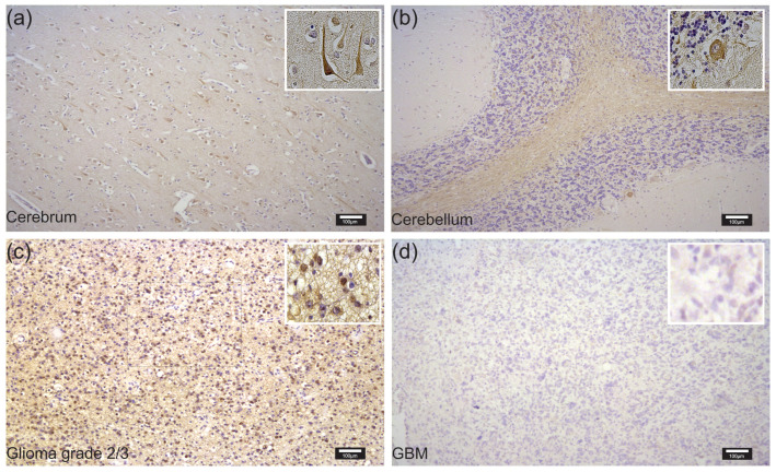
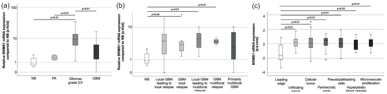
The metastatic suppressor BRMS1 interacts with critical steps of the metastatic cascade in many cancer entities. As gliomas rarely metastasize, BRMS1 has mainly been neglected in glioma research. However, its interaction partners, such as NFκB, VEGF, or MMPs, are old acquaintances in neurooncology. The steps regulated by BRMS1, such as invasion, migration, and apoptosis, are commonly dysregulated in gliomas. Therefore, BRMS1 shows potential as a regulator of glioma behavior. By bioinformatic analysis, in addition to our cohort of 118 specimens, we determined BRMS1 mRNA and protein expression as well as its correlation with the clinical course in astrocytomas IDH mutant, CNS WHO grade 2/3, and glioblastoma IDH wild-type, CNS WHO grade 4. Interestingly, we found BRMS1 protein expression to be significantly decreased in the aforementioned gliomas, while BRMS1 mRNA appeared to be overexpressed throughout. This dysregulation was independent of patients' characteristics or survival. The protein and mRNA expression differences cannot be finally explained at this stage. However, they suggest a post-transcriptional dysregulation that has been previously described in other cancer entities. Our analyses present the first data on BRMS1 expression in gliomas that can provide a starting point for further investigations.

Keywords: behavior; glioblastoma; mRNA; metastasis; protein; suppressor.


Conflict of interest statement

The authors declare no conflict of interest. The funders had no role in the design of the study, in the collection, analysis, or interpretation of data, in the writing of the manuscript, or in the decision to publish the results.

Figures

Figure 1
Figure 1
Involvement of BRMS1 in critical steps of the metastatic cascade. Adapted from [40] and created with BioRender.com. BRMS1 interacts with several steps of the metastatic cascade. Its most prominent impact on invasion appears to be through the attenuation of nuclear factor-kappa B (NFκB) function via the classical/canonical pathway [41,42]. One underlying mode of action lies in BRMS1′s ability to bind to the NFκB region of the urokinase plasminogen-activator (uPA) promoter, a downstream target of NFκB, thereby suppressing its (usually) NFκB-dependent gene expression. However, other mechanisms of interaction have also been investigated [42]. Furthermore, BRMS1 expression has been described as inhibiting migratory behavior. In glioma cells, among others, the expression of the Focal Adhesion Kinase (FAK) and Src proteins decreased when BRMS1 was re-expressed [36]. Cell culture experiments further suggest that BRMS1 may interact with VEGF, COX2, and MMPs to prevent cell adhesion and extravasation [42]. After arriving in their target tissue, the disseminated cells need to be able to exit dormancy and begin proliferation again to properly colonize and build a metastasis. BRMS1 can inhibit metastatic outgrowth and has been described as being associated with an increase in PARP and caspase-3 levels, though the molecular mechanisms behind this association have not been exhaustively understood [38,42].
Figure 2
Figure 2
Representative examples of immunohistochemical staining with DAB and a BRMS1 antibody in NB, glioma grade 2/3 and GBM tissue: (a) staining of healthy cerebrum; (b) healthy cerebellum; (c) glioma grade 2/3; and (d) GBM. All photographs were taken using the same settings. Magnifications are equal in all four pictures, and scale bars represent 100 µm.
Figure 3
Figure 3
BRMS1 mRNA expression in gliomas. (a) BRMS1 mRNA expression in gliomas compared to non-cancerous brain (NB: n = 12; PA: n = 4; gliomas grade 2/3: n = 24, GBM: n = 78; ANOVA: p < 0.05, Levene’s test: p < 0.05, post-hoc: Dunnet—T3). (b) GBM-subgroup analysis of tumors with different growth patterns (NB: n = 12; GBM primary tumor leading to local relapse: n = 24; GBM local relapse: n = 8; GBM primary tumor leading to multifocal relapse: n = 10; GBM multifocal relapse: n = 3; GBM multifocal primary tumor: n = 10; ANOVA: p > 0.05). (c) IVY-GAP database [46] analysis of BRMS1 mRNA expression in different areas of GBM (leading edge: n = 19; infiltrating tumor: n = 24; cellular tumor: n = 111; perinecrotic zone: n = 26; pseudopalisading cells around necrosis: n = 40; hyperplastic blood vessels in cellular tumor: n = 22; microvascular proliferation: n = 28). Circles represent outliers. Abbreviations: NB, non-cancerous brain; PA, adult pilocytic astrocytoma; GBM, glioblastoma.
Figure 4
Figure 4
Kaplan-Meier analyses of overall (OS) and progression-free survival (PFS) (a) OS (low BRMS1-mRNA expression: n = 15; intermediate BRMS1-mRNA expression: n = 14, high BRMS1-mRNA expression n = 15; Log Rank: p > 0.05) and PFS of GBM patients from our collective (low BRMS1-mRNA expression: n = 12; intermediate BRMS1-mRNA expression: n = 14, high BRMS1-mRNA expression n = 12; Log Rank: p > 0.05). Six patients died without matching the MRI-based RANO criteria for progress and therefore were excluded from the PFS analyses [53]. (b) OS (low BRMS1-mRNA expression: n = 42, intermediate BRMS1-mRNA expression: n = 65, high BRMS1-mRNA expression: n = 45, Log Rank: p > 0.05) and PFS of GBM patients from the TCGA dataset (low BRMS1-mRNA expression: n = 42, intermediate BRMS1-mRNA expression: n = 65, high BRMS1-mRNA expression: n = 45, Log Rank: p > 0.05). TCGA data were exported from the CBioportal analysis tool [54]. (c) OS of glioma grade 2/3 patients (low BRMS1-mRNA expression: n = 7; intermediate BRMS1-mRNA expression: n = 8; high BRMS1-mRNA expression n = 7; Log Rank: p > 0.05).

References

    1. Ostrom Q.T., Cioffi G., Waite K., Kruchko C., Barnholtz-Sloan J.S. CBTRUS Statistical Report: Primary Brain and Other Central Nervous System Tumors Diagnosed in the United States in 2014–2018. Neuro-Oncology. 2021;23:iii1–iii105. doi: 10.1093/neuonc/noab200. - DOI - PMC - PubMed
    1. Ostrom Q.T., Gittleman H., Truitt G., Boscia A., Kruchko C., Barnholtz-Sloan J.S. CBTRUS Statistical Report: Primary Brain and Other Central Nervous System Tumors Diagnosed in the United States in 2011–2015. Neuro-Oncology. 2018;20:iv1–iv86. doi: 10.1093/neuonc/noy131. - DOI - PMC - PubMed
    1. Stupp R., Mason W.P., van den Bent M.J., Weller M., Fisher B., Taphoorn M.J., Belanger K., Brandes A.A., Marosi C., Bogdahn U., et al. Radiotherapy plus concomitant and adjuvant temozolomide for glioblastoma. N. Engl. J. Med. 2005;352:987–996. doi: 10.1056/NEJMoa043330. - DOI - PubMed
    1. Wick W., Osswald M., Wick A., Winkler F. Treatment of glioblastoma in adults. Ther. Adv. Neurol. Disord. 2018;11:1756286418790452. doi: 10.1177/1756286418790452. - DOI - PMC - PubMed
    1. Wrensch M., Minn Y., Chew T., Bondy M., Berger M.S. Epidemiology of primary brain tumors: Current concepts and review of the literature. Neuro-Oncology. 2002;4:278–299. doi: 10.1093/neuonc/4.4.278. - DOI - PMC - PubMed