Review
. 2023 May 28;15(11):2953.
doi: 10.3390/cancers15112953.

Primary Ovarian Leiomyosarcoma Is a Very Rare Entity: A Narrative Review of the Literature

Affiliations
Review

Primary Ovarian Leiomyosarcoma Is a Very Rare Entity: A Narrative Review of the Literature

Vincenzo Dario Mandato et al. Cancers (Basel). .

Abstract

Background: Primary ovarian leiomyosarcoma is a very rare malignancy characterized by unclear management and poor survival. We reviewed all the cases of primary ovarian leiomyosarcoma to identify prognostic factors and the best treatment.

Methods: We collected and analyzed the articles published in the English literature regarding primary ovarian leiomyosarcoma from January 1951 to September 2022, using PubMed research. Clinical and pathological characteristics, different treatments and outcomes were analyzed.

Results: 113 cases of primary ovarian leiomyosarcoma were included. Most patients received surgical resection, associated with lymphadenectomy in 12.5% of cases. About 40% of patients received chemotherapy. Follow-up information was available for 100/113 (88.5%) patients. Stage and mitotic count were confirmed to affect survival, and lymphadenectomy and chemotherapy were associated with a better survival rate. A total of 43.4% of patients relapsed, and their mean disease-free survival was 12.5 months.

Conclusions: Primary ovarian leiomyosarcomas are more common in women in their 50s (mean age 53 years). Most of them are at an early stage at presentation. Advanced stage and mitotic count showed a detrimental effect on survival. Surgical excision associated with lymphadenectomy and chemotherapy are associated with increased survival. An international registry could help collect clear and reliable data to standardize the diagnosis and treatment.

Keywords: chemotherapy; lymphadenectomy; mitotic count; primary ovarian leiomyosarcoma; review; stage; survival; symptoms; treatment.


Conflict of interest statement

The authors declare no conflict of interest. Vincenzo Dario Mandato, Federica Torricelli, Valentina Mastrofilippo, Andrea Palicelli, Luigi Costagliola, Lorenzo Aguzzoli.

Figures

Figure 1
Figure 1
Review of the literature: Preferred Reporting Items for Systematic Reviews and Meta-Analyses (PRISMA) flow chart.
Figure 2
Figure 2
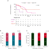
Impact of stage (A) and mitotic count (B) on OS of patients with POLMS.
Figure 3
Figure 3
Impact of surgery on OS of patients with POLMS (A). Impact of tumor stage on the choice of the treatment of POLMS patients (B). Impact of mitotic count on the choice of the treatment of POLMS patients (C).
Figure 4
Figure 4
Impact of the different treatments on OS (p = 0.016, Figure 4A) and on the risk of death (p = 0.032, Figure 4B) of POLMS patients. Impact of stage (C) and mitotic count (D) on the choice of treatment of POLMS patients. Impact of stage (E) and mitotic count (F) on the risk of death in POLMS patients.
Figure 5
Figure 5
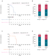
Impact of lymphadenectomy on OS of POLMS patients treated with chemotherapy (A). Impact of chemotherapy on OS of POLMS patients treated with lymphadenectomy (C). Impact of stage on the choice of chemotherapy (B) and lymphadenectomy (D).
Figure 6
Figure 6
Ultrasound appearance of POLMS: a huge pelvic mass of 20 × 15 × 16 cm occupying the pelvis going beyond the transverse umbilical, it appears as a solid mass with irregular margins (red arrow), dishomogeneous echostructure due to the presence of anechoic, poorly vascularized cystic areas at color doppler (yellow star) (previously unpublished, original photos).
Figure 7
Figure 7
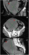
Computed tomography appearance of POLMS: Coronal (A), Axial (B) and Sagittal (C) plane: gross expansive lesion originating from the right adnexa adhering to the right wall of the uterus. It presents a mixed-cystic solid structure with a central fluid component (yellow star) and multiple solid peripheral vegetations (red arrow) with contrastographic enhancement (previously unpublished, original photos).
Figure 8
Figure 8
The tumor revealed a proliferation of spindle cells with hyperchromasia and moderate-severe nuclear atypia. The tumor borders were predominantly pushing with a residual peripheral focus of ovarian parenchyma (1A, star). Mitotic figures were frequently identified (1B, arrows) (A): Hematoxylin and eosin, 20 ×; (B): Hematoxylin and eosin, 10 ×)) (previously unpublished, original photos).
Figure 9
Figure 9
Histological details of the tumor cells. Spindle cell morphology. Severe pleomorphism. Necrotic areas (2B, star) (hematoxylin and eosin; (A): 40 ×; (B): 20 ×; (C): 40 ×). (D) On immunohistochemical exam, the tumor cells were positive for smooth muscle actin (20 ×) (previously unpublished, original photos).

References

    1. Arslan O.S., Sumer C., Cihangiroğlu G., Kanat-Pektas M., Gungor T. A rare tumor of the female genital tract: Primary ovarian leiomyosarcoma. Arch. Gynecol. Obstet. 2010;283:83–85. doi: 10.1007/s00404-010-1735-3. - DOI - PubMed
    1. Cojocaru E., Gamage G.P., Butler J., Barton D.P., Thway K., Fisher C., Messiou C., Miah A.B., Zaidi S., Gennatas S., et al. Clinical management and outcomes of primary ovarian leiomyosarcoma—Experience from a sarcoma specialist unit. Gynecol. Oncol. Rep. 2021;36:100737. doi: 10.1016/j.gore.2021.100737. - DOI - PMC - PubMed
    1. Kozłowski M., Nowak K., Kordek A., Cymbaluk-Płoska A. Therapeutic Management of Rare Primary Ovarian Neoplasms: Carcinosarcoma, Leiomyosarcoma, Melanoma and Carcinoid. Int. J. Environ. Res. Public Health. 2021;18:7819. doi: 10.3390/ijerph18157819. - DOI - PMC - PubMed
    1. Goodall E.J., Madhuri T., Manuel S.B. The Management Dilemma of Leiomyosarcoma of the Ovary. World J. Oncol. 2011;2:265–266. doi: 10.4021/wjon362w. - DOI - PMC - PubMed
    1. Inoue J., Gomibuchi H., Minoura S. A Case of a Primary Ovarian Leiomyosarcoma. J. Obstet. Gynaecol. Res. 2000;26:401–407. doi: 10.1111/j.1447-0756.2000.tb01349.x. - DOI - PubMed

LinkOut - more resources