. 2023 May 30;15(11):2987.
doi: 10.3390/cancers15112987.

Matrix Metalloproteinase 9 Induced in Esophageal Squamous Cell Carcinoma Cells via Close Contact with Tumor-Associated Macrophages Contributes to Cancer Progression and Poor Prognosis

Affiliations

Matrix Metalloproteinase 9 Induced in Esophageal Squamous Cell Carcinoma Cells via Close Contact with Tumor-Associated Macrophages Contributes to Cancer Progression and Poor Prognosis

Shuichi Tsukamoto et al. Cancers (Basel). .

Abstract

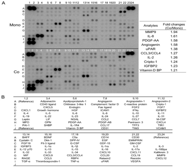
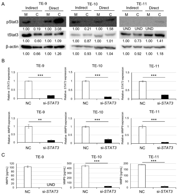
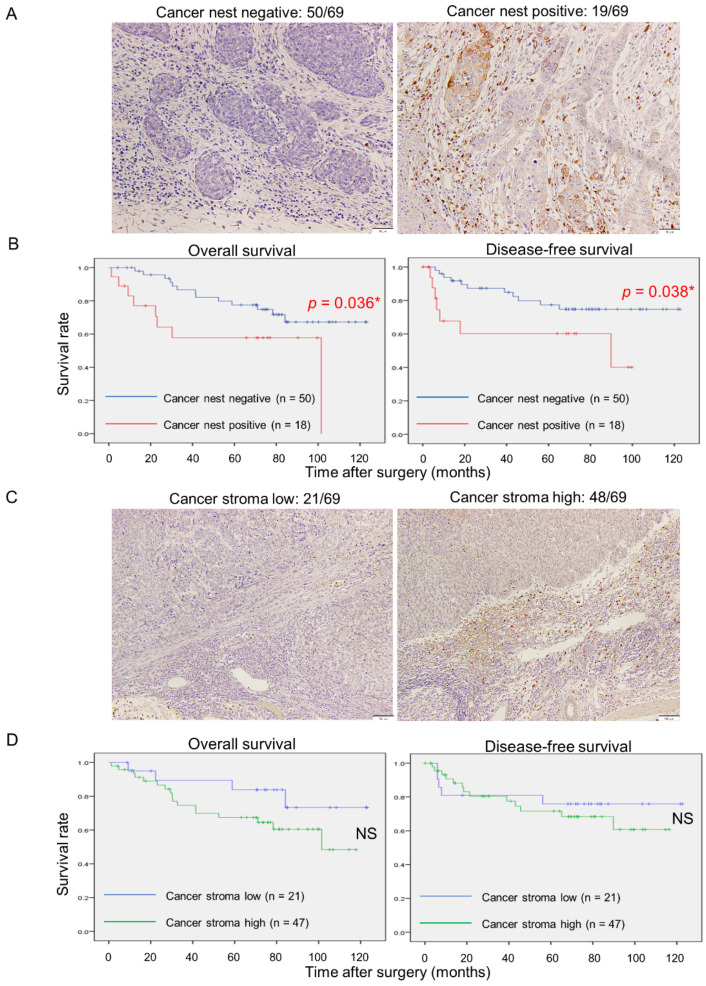
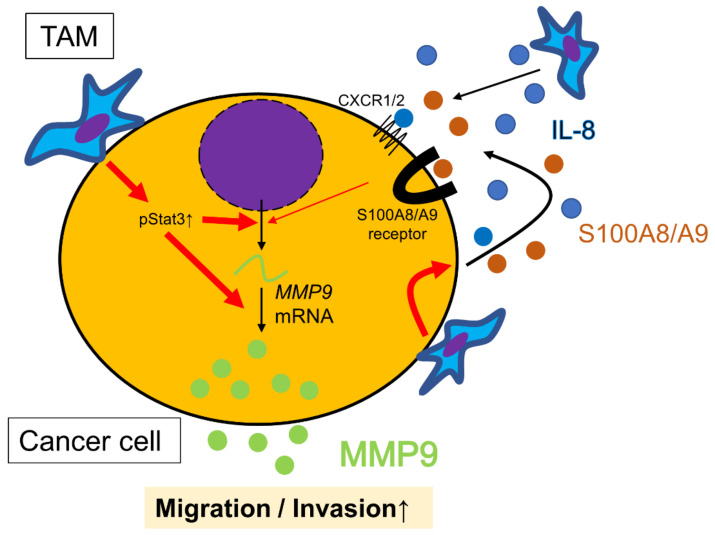
Tumor-associated macrophages (TAMs) contribute to disease progression in various cancers, including esophageal squamous cell carcinoma (ESCC). We have previously used an indirect co-culture system between ESCC cell lines and macrophages to analyze their interactions. Recently, we established a direct co-culture system to closely simulate actual ESCC cell-TAM contact. We found that matrix metalloproteinase 9 (MMP9) was induced in ESCC cells by direct co-culture with TAMs, not by indirect co-culture. MMP9 was associated with ESCC cell migration and invasion, and its expression was controlled by the Stat3 signaling pathway in vitro. Immunohistochemical analyses revealed that MMP9 expression in cancer cells at the invasive front ("cancer cell MMP9") was related to high infiltration of CD204 positive M2-like TAMs (p < 0.001) and was associated with worse overall and disease-free survival of patients (p = 0.036 and p = 0.038, respectively). Furthermore, cancer cell MMP9 was an independent prognostic factor for disease-free survival. Notably, MMP9 expression in cancer stroma was not associated with any clinicopathological factors or patient prognoses. Our results suggest that close interaction with TAMs infiltrating in cancer stroma or cancer nests induces MMP9 expression in ESCC cells, equipping them with more malignant features.

Keywords: activator of transcription 3; direct co-culture; direct contact; esophageal squamous cell carcinoma; immunohistochemistry; interleukin-8; matrix metalloproteinase 9; prognostic factor; signal transducer; tumor-associated macrophage.


Conflict of interest statement

The authors declare no conflict of interest.

Figures

Figure 1
Figure 1
Cytokine array analysis shows increased MMP9 secretion from TE-11 cells after direct co-culture with macrophages. (A): Cytokine array analysis of culture supernatants from mono- (upper array) and co-cultured (lower array) TE-11 cells. The densities of spots were analyzed using the ImageJ 1.53t and the top 10 increased fold changes in densities are listed on the right. (B): Coordinates of the cytokine array.
Figure 2
Figure 2
Direct co-culture with macrophages induces MMP9 gene expression and protein secretion in ESCC cells, whereas indirect co-culture does not. (A): qPCR revealed upregulation of MMP9 gene transcription in ESCC cells by direct co-culture with macrophages but not by indirect co-culture. (B): ELISA quantified MMP9 in culture supernatants of mono- and indirectly or directly co-cultured ESCC cell lines. MMP9 secretion from ESCC cells was elevated by the direct co-culture but not by the indirect co-culture, except for TE-10. (C): Gelatin zymography showed increased MMP9 secretion by ESCC cells directly co-cultured with macrophages, and the gelatinolytic activity of MMP9 was preserved in the culture supernatants. Band densities were calculated using the ImageJ 1.53t and expressed as relative values. The band density of monocultured conditions in each cell line was set as 1.00. The indirect co-culture system was performed in 6-well plates, while the direct co-culture system was conducted in 10-cm dishes. Data are exhibited as mean ± SEM; * p < 0.05, ** p < 0.01, *** p < 0.001; M, monocultured; C, co-cultured; UND, undetected.
Figure 3
Figure 3
Direct co-culture with macrophages triggers Stat3 phosphorylation, and Stat3 signal pathway may contribute to MMP9 upregulation in ESCC cells. (A): Western blot for phosphorylated (p) Stat3 demonstrated increased Stat3 phosphorylation by the direct co-culture but not by the indirect co-culture. (B): qPCR showed that STAT3 and MMP9 gene transcriptions in ESCC cells were significantly suppressed by STAT3 knockdown. (C): ELISA determined that MMP9 protein secretion from ESCC cells was significantly downregulated by STAT3 knockdown. Band densities were calculated using the ImageJ 1.53t and expressed as relative values. The band density of monocultured conditions in each cell line was set as 1.00. pStat3 was not detected in TE-11 cells of monocultured counterparts and after the indirect co-culture. pStat3, phosphorylated-Stat3; tStat3, total-Stat3; M, monocultured; C, co-cultured; NC, siRNA of negative control; si-STAT3, siRNA against STAT3; UND, undetected. Data are presented as mean ± SEM; UND: undetected; ** p < 0.01, *** p < 0.001.
Figure 4
Figure 4
Direct co-culture with macrophages enhances migration and invasion of ESCC cells, which is abrogated by an MMP9 Inhibitor. (A): Transwell migration assays revealed that the direct co-culture increased ESCC cell migration. Moreover, the increase was nullified by the MMP9 inhibitor ab142180, except in the assay using TE-11. (B): Representative images in the transwell migration assay of TE-11. (C): Transwell invasion assays showed that the direct co-culture increased ESCC cell invasion. As in migration assays, increased invasion was significantly abrogated by ab142180 in the assays of TE-9 and TE-11 but not in TE-10, although an inhibitory trend was observed. (D): Representative images in the transwell invasion assay of TE-11. Data are exhibited as mean ± SEM; * p < 0.05, ** p < 0.01, *** p < 0.001.
Figure 5
Figure 5
MMP9 immunoreactivity of cancer cells at the invasive front, but not stromal cells, in human ESCC tissues significantly correlates with poorer clinical outcomes. (A): Representative images of MMP9 IHC at the invasive front of ESCC tissues. Positive staining was defined as stronger staining than surrounding fibroblasts or lymphocytes. Left, negative (50 out of total 69 cases); Right, positive (19 out of total 69 cases). Scale bars: 50 μm. (B): Kaplan–Meier survival analyses of overall (Left) and disease-free survival (Right) based on MMP9 expression of cancer cells at the invasive front. (C): Representative images of MMP9 IHC in the cancer stroma of ESCC tissues. Stromal MMP9 expression was classified as low (Left, 21 out of total 69 cases) or high (Right, 48 out of total 69 cases) expressions based on the intensity of adjacent non-neoplastic tissue. Scale bars: 100 μm. (D): Kaplan–Meier survival analyses of overall (Left) and disease-free survival (Right) based on MMP9 expression of cancer stromal cells. Data were compared with the log-rank test; * p < 0.05; NS, not significant.
Figure 6
Figure 6
IL-8 as a partial inducer of MMP9 in ESCC cells. (A): ELISA quantification of IL-8 in culture supernatants of ESCC cells after direct co-culture with macrophages. Directly co-cultured ESCC cells secreted more IL-8 than monocultured counterparts. (B): Western blot revealed the expression of known IL-8 receptors, CXCR1 (45 kDa, monomer; 80–90 kDa, dimer and their glycosylated forms) and CXCR2 (41 kDa) in TE-9, TE-10, and TE-11 cell lines. (C,D): Treatment with rhIL-8 (100 ng/mL, for 24 h) upregulated MMP9 mRNA expression in all three ESCC cell lines (C) but only triggered MMP9 secretion from TE-10 (D). Data are expressed as mean ± SEM; * p < 0.05, ** p < 0.01, *** p < 0.001. NS, not significant.
Figure 7
Figure 7
Schematic model of the interactions between ESCC cells and tumor-associated macrophages (TAMs) from the viewpoint of MMP9 regulation in ESCC cells. Direct contact between ESCC cells and TAMs strongly upregulates MMP9 transcription and secretion, promoting migration and invasion of ESCC cells. TAMs also release IL-8, which indirectly induces MMP9 gene transcription. In addition, the direct contact between ESCC cells and TAMs induces the secretion of IL-8 and S100A8/A9 from ESCC cells, contributing to their migration, invasion and MMP9 production in autocrine or paracrine manner. Collectively, direct contact more effectively promotes ESCC cell motility and invasion than indirect interaction alone.

Similar articles

Cited by

References

    1. Sheikh M., Roshandel G., McCormack V., Malekzadeh R. Current Status and Future Prospects for Esophageal Cancer. Cancers. 2023;15:765. doi: 10.3390/cancers15030765. - DOI - PMC - PubMed
    1. Cancer Statistics Cancer Information Service, National Cancer Center, Japan (National Cancer Registry, Ministry of Health, Labour and Welfare) [(accessed on 24 February 2023)]. Available online: https://ganjoho.jp/reg_stat/statistics/data/dl/index.html#a14.
    1. Watanabe M., Toh Y., Ishihara R., Kono K., Matsubara H., Miyazaki T., Morita M., Murakami K., Muro K., Numasaki H., et al. Comprehensive registry of esophageal cancer in Japan, 2015. Esophagus Off. J. Jpn. Esophageal Soc. 2023;20:1–28. doi: 10.1007/s10388-022-00950-5. - DOI - PMC - PubMed
    1. Shimada H., Nabeya Y., Okazumi S.-I., Matsubara H., Shiratori T., Gunji Y., Kobayashi S., Hayashi H., Ochiai T. Prediction of survival with squamous cell carcinoma antigen in patients with resectable esophageal squamous cell carcinoma. Surgery. 2003;133:486–494. doi: 10.1067/msy.2003.139. - DOI - PubMed
    1. Komohara Y., Fujiwara Y., Ohnishi K., Takeya M. Tumor-associated macrophages: Potential therapeutic targets for anti-cancer therapy. Adv. Drug Deliv. Rev. 2016;99:180–185. doi: 10.1016/j.addr.2015.11.009. - DOI - PubMed

LinkOut - more resources