. 2023 May 30;16(11):4080.
doi: 10.3390/ma16114080.

Biocompatibility, Surface Morphology, and Bacterial Load of Dental Implant Abutments following Decontamination Protocols: An In-Vitro Study

Affiliations

Biocompatibility, Surface Morphology, and Bacterial Load of Dental Implant Abutments following Decontamination Protocols: An In-Vitro Study

Esi Sharon et al. Materials (Basel). .

Abstract

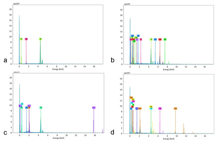
The long-term success of dental implant rehabilitation depends significantly on proper peri-implant soft tissue integration. Therefore, decontamination of abutments prior to their connection to the implant is beneficial to enhance soft tissue attachment and to aid in maintaining marginal bone around the implant. Consequently, different implant abutment decontamination protocols were evaluated regarding biocompatibility, surface morphology, and bacterial load. The protocols evaluated were autoclave sterilization, ultrasonic washing, steam cleaning, chlorhexidine chemical decontamination, and sodium hypochlorite chemical decontamination. The control groups included: (1) implant abutments prepared and polished in a dental lab without decontamination and (2) unprepared implant abutments obtained directly from the company. Surface analysis was performed using scanning electron microscopy (SEM). Biocompatibility was evaluated using XTT cell viability and proliferation assays. Biofilm biomass and viable counts (CFU/mL) (n = 5 for each test) were used for surface bacterial load evaluation. Surface analysis revealed areas of debris and accumulation of materials, such as iron, cobalt, chromium, and other metals, in all abutments prepared by the lab and with all decontamination protocols. Steam cleaning was the most efficient method for reducing contamination. Chlorhexidine and sodium hypochlorite left residual materials on the abutments. XTT results showed that the chlorhexidine group (M = 0.7005, SD = 0.2995) had the lowest values (p < 0.001) (autoclave: M = 3.6354, SD = 0.1510; ultrasonic: M = 3.4077, SD = 0.3730; steam: M = 3.2903, SD = 0.2172; NaOCl: M = 3.5377, SD = 0.0927; prep non-decont.: M = 3.4815, SD = 0.2326; factory: M = 3.6173, SD = 0.0392). Bacterial growth (CFU/mL) was high in the abutments treated with steam cleaning and ultrasonic bath: 2.93 × 10⁹, SD = 1.68 × 10¹² and 1.83 × 10⁹, SD = 3.95 × 10¹⁰, respectively. Abutments treated with chlorhexidine showed higher toxicity to cells, while all other samples showed similar effects to the control. In conclusion, steam cleaning seemed to be the most efficient method for reducing debris and metallic contamination. Bacterial load can be reduced using autoclaving, chlorhexidine, and NaOCl.

Keywords: decontamination; osseointegration; peri-implantitis; steam; titanium; ultrasonics.


Conflict of interest statement

The authors declare no conflict of interest.

Figures

Figure 1
Figure 1
Factory abutments that did not undergo laboratory preparation ((a) no magnification; (b) ×60 magnification; (c) ×500 magnification; (d) ×1000 magnification; (e) ×2000 magnification). The samples are clean, smooth, and devoid of any signs of debris and/or contamination.
Figure 2
Figure 2
Abutments that were prepared in a lab but did not undergo any decontamination method ((a) no magnification; (b) ×60 magnification; (c) ×500 magnification; (d) ×1000 magnification; (e) ×2000 magnification). Apart from the noticeable scratches caused by the tungsten carbide burs used to prepare the abutments, the samples are covered with various debris and contaminants.
Figure 3
Figure 3
Abutments that were prepared in a lab and decontaminated using steam cleaning ((a) no magnification; (b) ×60 magnification; (c) ×500 magnification; (d) ×1000 magnification; (e) ×2000 magnification). The samples have some debris, but to a lesser extent than the control samples in Figure 2 and look smoother.
Figure 4
Figure 4
Abutments that were prepared in a lab and decontaminated using ultrasonic washing ((a) no magnification; (b) ×60 magnification; (c) ×500 magnification; (d) ×1000 magnification; (e) ×2000 magnification). The samples have some debris and accumulation of contaminants, but look smoother compared to the control samples shown in Figure 2.
Figure 5
Figure 5
Abutment that was prepared in a lab and decontaminated using autoclave sterilization ((a) no magnification; (b) ×60 magnification; (c) ×500 magnification; (d) ×1000 magnification; (e) ×2000 magnification). The sample has some debris and accumulation of contaminants, but to a lesser extent than the control samples in Figure 2.
Figure 6
Figure 6
Abutment that was prepared in a lab and decontaminated using chlorhexidine ((a) no magnification; (b) ×60 magnification; (c) ×500 magnification; (d) ×1000 magnification; (e) ×2000 magnification). The sample has some debris and accumulation of contaminants, but to a lesser extent than the control samples in Figure 2. In addition, the sample is covered with residual material from the CHX solution.
Figure 7
Figure 7
Abutment that was prepared in a lab and decontaminated using sodium hypochlorite ((a) no magnification; (b) ×60 magnification; (c) ×500 magnification; (d) ×1000 magnification; (e) ×2000 magnification). It is noticeable that, in comparison to the control samples seen in Figure 2, the sample is covered with major residual material from the NaOCl solution, as well as pits and flaws apparently caused by the sodium hypochlorite solution.
Figure 8
Figure 8
Chemical analysis of abutments under different treatments. The different peaks represent various metallic contaminations that were analyzed on the samples. (a) abutments from factory, (b) abutments after lab preparation, (c) abutments after lab preparation and steam cleaning, and (d) abutments after lab preparation and ultrasonic washing.
Figure 9
Figure 9
XTT assay results. The various decontamination methods were assessed (autoclave, ultrasonic washing, steam cleaning (steam), chlorhexidine (CHX), and sodium hypochlorite (NaOCl)), as well as 5 abutments that were prepared in a lab but did not undergo any decontamination method (prep non-decont.) and 5 abutments from the factory that did not undergo any decontamination method (factory). Additional control groups included macrophage-like raw cells cultured in medium (cells), as well as supplemented with DMEM (medium). The average XTT values of CHX were significantly lower than the XTT values of any of the other groups and close to the values of the supplemental DMEM (medium). The rest of the groups showed XTT values similar to each another.
Figure 10
Figure 10
Comparison of mean bacterial counts (CFU/mL) following decontamination procedures. Groups 1 and 2 (controls: unprepared untreated abutments and prepared untreated abutments). Groups 3–9 abutments after different decontamination procedures. No bacterial growth was observed in the implant abutments treated with autoclave, chlorhexidine, chlorhexidine + saline, NaOCl solution, and NaOCl solution + saline. Very low bacterial growth was seen in implant abutments treated with chlorhexidine. Abutments treated with steam cleaning and ultrasonic bath showed bacterial growth closely resembling the control groups.

Similar articles

References

    1. Pjetursson B.E., Sailer I., Latyshev A., Rabel K., Kohal R.J., Karasan D. A systematic review and meta-analysis evaluating the survival, the failure, and the complication rates of veneered and monolithic all-ceramic implant-supported single crowns. Clin. Oral Implants Res. 2021;32((Suppl. S21)):254–288. doi: 10.1111/clr.13863. - DOI - PMC - PubMed
    1. Pjetursson B.E., Thoma D., Jung R., Zwahlen M., Zembic A. A systematic review of the survival and complication rates of implant-supported fixed dental prostheses (FDPs) after a mean observation period of at least 5 years. Clin. Oral Implants Res. 2012;23((Suppl. S6)):22–38. doi: 10.1111/j.1600-0501.2012.02546.x. - DOI - PubMed
    1. Jung R.E., Pjetursson B.E., Glauser R., Zembic A., Zwahlen M., Lang N.P. A systematic review of the 5-year survival and complication rates of implant-supported single crowns. Clin. Oral Implants Res. 2008;19:119–130. doi: 10.1111/j.1600-0501.2007.01453.x. - DOI - PubMed
    1. Jung R.E., Zembic A., Pjetursson B.E., Zwahlen M., Thoma D.S. Systematic review of the survival rate and the incidence of biological, technical, and aesthetic complications of single crowns on implants reported in longitudinal studies with a mean follow-up of 5 years. Clin. Oral Implants Res. 2012;23((Suppl. S6)):2–21. doi: 10.1111/j.1600-0501.2012.02547.x. - DOI - PubMed
    1. Welander M., Abrahamsson I., Berglundh T. The mucosal barrier at implant abutments of different materials. Clin. Oral Implants Res. 2008;19:635–641. - PubMed

LinkOut - more resources