. 2023 May 23;12(11):3623.
doi: 10.3390/jcm12113623.

Unravelling the Clinical Co-Morbidity and Risk Factors Associated with Agenesis of the Corpus Callosum

Affiliations

Unravelling the Clinical Co-Morbidity and Risk Factors Associated with Agenesis of the Corpus Callosum

Callum J Smith et al. J Clin Med. .

Abstract

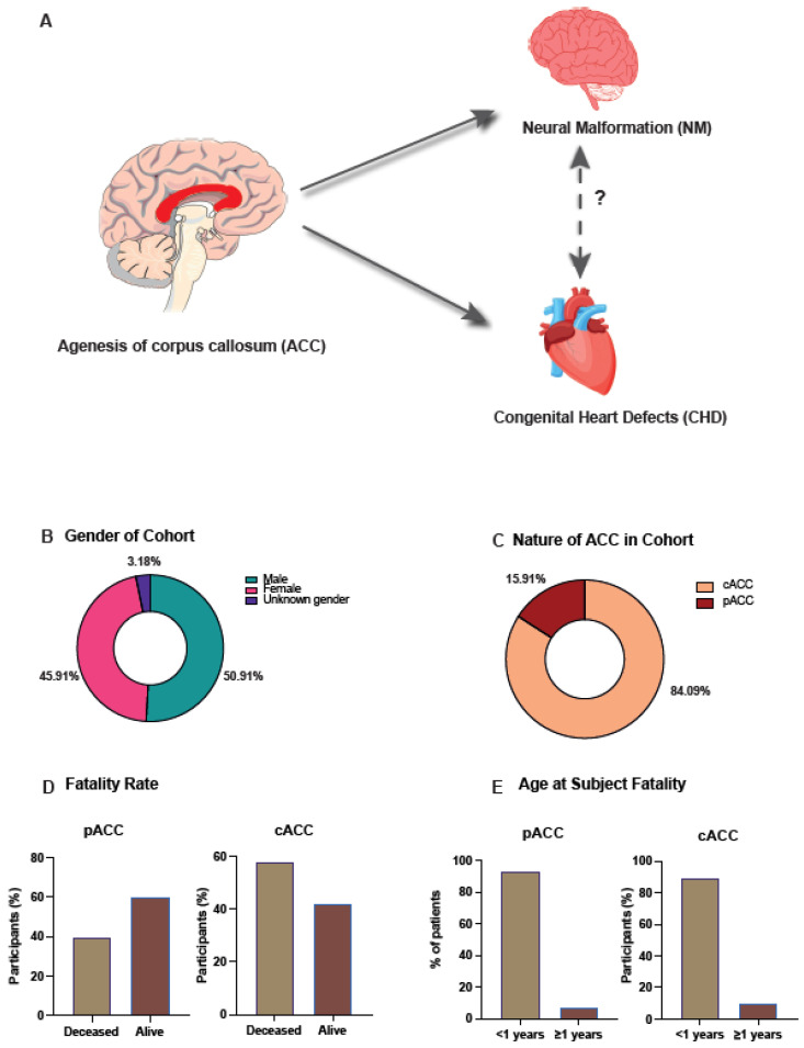
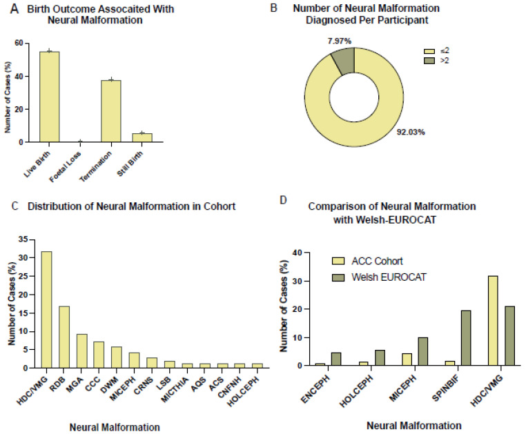
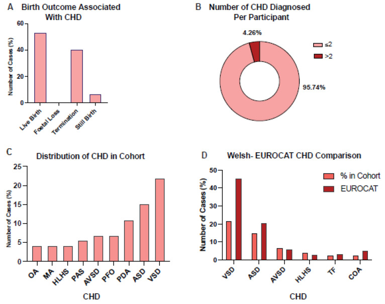
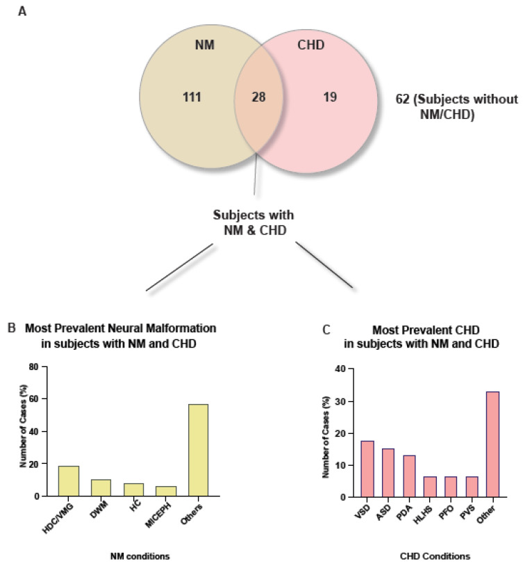
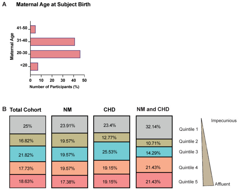
Agenesis of the Corpus Callosum (ACC) can result in multiple neurological deficits including social and behavioural issues. However, the underlying aetiology, clinical co-morbidity and the contributing risk factors remain elusive, resulting in inaccurate prognosis and delayed therapy. The main objective of this study was to comprehensively describe the epidemiology and clinical co-morbidity associated with patients diagnosed with ACC. The secondary objective was to identify the factors that contribute towards increased risk for ACC. For this, we analysed 22 years (1998-2020) of clinical data across the whole of Wales, UK collected through the Congenital Anomaly Register & Information Service (CARIS) and Public Health Wales (PHW). Our results demonstrate that complete ACC (84.1%) was the prevalent subtype, in comparison to partial ACC. Further, ventriculomegaly/hydrocephalus (26.37%) and ventricular septal defect (21.92%) were identified to be the most prevalent neural malformation (NM) and congenital heart disorder (CHD) in our cohort. Although 12.7% of subjects with ACC had both an NM and CHD, we found no significant association between them (χ2 (1, n = 220) = 3.84, p = 0.33). We found socioeconomic deprivation and increased maternal age contributed towards an increased risk for ACC. To the best of our knowledge, this study for the first time defines the clinical phenotypes and the factors that contribute to ACC within the Welsh population. These findings will be of value to both patients and healthcare professionals, who may take preventative or remedial measures.

Keywords: agenesis of corpus callosum; comorbidity; congenital heart disorders; neurodevelopmental disorders; risk factors.


Conflict of interest statement

All other authors declared no conflict of interest.

Figures

Figure 1
Figure 1
Graphical abstract and graphs depicting the demographics of study participants. (A) One of the aims of the study was to analyse whether there is an association between neural malformations (NM) and congenital heart defects (CHD) amongst those diagnosed with agenesis of the corpus callosum (ACC) as associations between ACC and NM and ACC and CHD have already been established. (B) Raw data from 220 patients with ACC provided by Public Health Wales was filtered and the data shows that 45.91% (n = 101) of those with ACC between 1998–2020 were female, 50.91% (n = 112) were male and 3.18% (n = 7) had unknown gender due to either foetal loss or termination of the pregnancy occurring before gender could be determined. (C) The ACC morphology of the cohort was determined with 84.09% (n = 185) showing complete ACC (cACC) and 15.91% (n = 35) showing partial ACC (pACC). (D) The fatality rate of the cohort was calculated and subsequently broken down to show the rate of fatality within each ACC morphology group. Of those with pACC 40% (n = 14) had passed away when the study was conducted and 60% (n = 21) were alive. In contrast, of those with cACC 57.84% (n = 107) had passed away and 42.16% (n = 78) were alive. (E) To further understand the demographics surrounding fatality of individuals with ACC the age at which the participant died was explored. 92.86% (n = 13) of those diagnosed with pACC died at <1 year of age. Similarly, 89.72% (n = 96) of the cACC cohort who had passed away died at <1 year of age.
Figure 2
Figure 2
Graphs depicting the demographics of those with ACC that also had neural malformation (NM) diagnoses. (A) The birth outcome of those with ACC and NM (n = 138) was explored to investigate whether having an NM diagnosis in addition to the ACC diagnosis affected birth outcome. The data shows that live birth was the most common outcome for this cohort (55.8%, n = 77), followed by termination of pregnancy (37.68%, n = 52), then stillbirth (5.8%, n = 8) and lastly foetal loss (0.72%, n = 1). (B) The pie chart shows the demographics of NM diagnosis in this study’s cohort. 92.03% (n = 127) had two or less NM diagnoses, whilst 7.97% (n = 11) had more than two NM diagnoses. (C) The bar chart is showing which NM diagnoses were identified in this cohort and how prevalent they were amongst this cohort. Ventriculomegaly/hydrocephalus (HDC/VMG) (26.37%, n = 53) was the most common NM, followed by reduction deformities of the brain (ROB) (16.92%, n = 34), microgyria (MGA) (9.45%, n = 19), congenital cerebral cysts (CCC) (7.46%, n = 15), and Dandy–Walker malformation (DWM) (5.47%, n = 11). Other NM diagnoses were colpocephaly (COLCEPH) (5.47%, n = 11), microcephaly (MICEPH) (4.48%, n = 9), craniosynostosis (CRNS) (2.99%, n = 6), lumbar spina bifida (LSB) (1.99%, n = 4), micrognathia (MICTHIA) (1.49%, n = 3), aqueduct Stenosis (AQS) (1.49%, n = 3), Arnold–Chiari syndrome (ACS) (1.49%, n = 3), congenital malformation of face and neck, unspecified (CNFNH) (1.49%, n = 3) and holoprosencephaly (HOLCEPH) (1.49%, n = 3). (D) To understand whether NM occurred more or less frequently among those with ACC compared to those with ACC, the prevalence of NM among this cohort was compared to data from EUROCAT (European network of population-based registries for the epidemiological surveillance of congenital anomalies). This analysis showed that this cohort of individuals with ACC had a 3.56% higher prevalence of HDC/VMG than the EUROCAT cohort although this was not statistically significant. Spinal bifida (SPINBIF) (p = 4 × 10⁻⁶), microcephaly (MICEPH) (p = 0.0036), holoprosencephaly (HOLOCEPH) (p = 0.0055), and encephalocele (ENCEPH) (p = 0.0069) prevalence was significantly higher in the EUROCAT population.
Figure 3
Figure 3
Graphs depicting the demographics of those with ACC that also had congenital heart defects (CHD) diagnoses. (A) The birth outcome of those with ACC and CHD (n = 47) was explored to investigate whether having a CHD diagnosis in addition to the ACC diagnosis affected birth outcome. The data shows that live birth was the most common outcome for this cohort (53.19%, n = 25), followed by termination of pregnancy (40.43%, n = 19), then stillbirth (6.38%, n = 3) and lastly foetal loss (0%, n = 0). (B) The pie chart shows the demographics of CHD diagnosis in this study’s cohort. 95.74% (n = 45) had less than or two NM diagnoses, whilst 4.26% (n = 2) had more than two CHD diagnoses. (C) The bar chart is showing which CHD diagnoses were made in this cohort and how prevalent they were amongst this cohort. Ventricular septal defect (VSD) was the most common CHD (21.92%, n = 16), followed by atrial septal defect (ASD) (15.07%, n = 11), and patent ductus arteriosum (PDA) (10.96%, n = 8). Other CHD diagnoses were, patent foramen ovale (PFO) (6.85%, n = 5), atrioventricular septal defect (AVSD) (6.85%, n = 5), pulmonary artery stenosis (PAS) (5.48%, n = 4), hypoplastic left heart syndrome (HLHS) (4.11%, n = 3), malformation of aorta (MA) (4.11%, n = 3) and overriding aorta (OA) (4.11%, n = 3). (D) To understand whether CHD occurred more or less frequently among those with ACC compared to those with ACC, the prevalence of CHD among this cohort was compared to data from EUROCAT (European network of population-based registries for the epidemiological surveillance of congenital anomalies). This analysis showed that the EUROCAT cohort had a significantly greater prevalence of VSD compared to our cohort (p = 4.7 × 10⁻⁵). Although, our cohort did have a significantly greater prevalence of PDA compared to EUROCAT data (p = 0.011). The CHDs compared were, ventricular septal defect (VSD), atrial septal defect (ASD), Atrioventricular Septal Defect (AVSD), Hypoplastic Left Heart Syndrome (HLHS), Tetralogy of Fallot (TF) and Coarctation of Aorta (COA).
Figure 4
Figure 4
Pie chart showing the demographics of diagnoses among those with ACC and bar charts depicting the most prevalent diagnoses among those with both an NM and CHD diagnosis. (A) The pie chart is showing the breakdown of diagnoses across this cohort and therefore the rate of co-morbidity among the cohort. All subjects have an ACC diagnosis, with 50.45% (n = 111) also having at least one NM diagnosis, 8.63% (n = 19) having at least one CHD diagnosis, 28.18% (n = 62) only having an ACC diagnosis and 25.54% (n = 28) having both at least one NM and CHD diagnosis in addition to the ACC diagnosis. (B) The bar chart shows that among those with both an NM and CHD diagnosis HDC/VMG was the most frequently observed NM diagnosis (26.37%, n = 53). Other NM diagnoses were Dandy–Walker Malformation (DWM) (5.97%, n = 12), hypoplastic cerebellum (HC) (3.98%, n = 8), and microcephaly (MICEPH) (4.48%, n = 9). (C) The bar chart highlights that the most frequently diagnosed CHD, among those with an NM and CHD diagnosis, was ventral septal defects (VSD) (21.92%, n = 16). Other CHD diagnoses were atrial septal defects (ASD) (15.07%, n = 11), patent ductus arteriosum (PDA) (10.96%, n = 8), hypoplastic left heart syndrome (HLHS) (4.11%, n = 3), patent foramen ovale (PFO) (6.85%, n = 5) and pulmonary valve stenosis (PVS) (1.37%, n = 1).
Figure 5
Figure 5
Maternal age at subject birth and socioeconomic background demographics. (A) One of the aims was the investigate whether maternal age is a risk factor for an individual developing ACC. As such maternal age at subject birth was explored, and the bar chart shows that the 20–30 years of age range was the most common maternal age during birth, 46.36% (n = 102), closely followed by the 31–40 years range, 41.36% (n = 91), then <20 years of age, 7.27% (n = 16) and finally 41–50 years of age, 5% (n = 11). (B) To further explore potential risk factors for developing ACC the WIMD 2019 quintiles of deprivation were analysed to determine if the socioeconomic background of the participants mother is associated with the development of ACC and other co-morbidities. Quintile 1, the most deprived quintile, was the most observed quintile in the total cohort (25%, n = 55), only NM diagnosed cohort (23.91%, n = 33), and both NM and CHD cohort (32.14%, n = 9). A chi-squared test was also conducted to determine if there is a statistically significant association between the co-morbidities diagnosed (i.e., only NM, only CHD and, NM and CHD) and the WIMD 2019 quintile. No statistically significant association (p = 0.45) was found between the deprivation quintiles and the co-morbidities diagnosed.

Similar articles

References

    1. Schell-Apacik C.C., Wagner K., Bihler M., Ertl-Wagner B., Heinrich U., Klopocki E., Kalscheuer V.M., Muenke M., von Voss H. Agenesis and dysgenesis of the corpus callosum: Clinical, genetic and neuroimaging findings in a series of 41 patients. Am. J. Med. Genet. A. 2008;146A:2501–2511. doi: 10.1002/ajmg.a.32476. - DOI - PMC - PubMed
    1. Hofman J., Hutny M., Sztuba K., Paprocka J. Corpus callosum agenesis: An insight into the etiology and spectrum of symptoms. Brain Sci. 2020;10:625. doi: 10.3390/brainsci10090625. - DOI - PMC - PubMed
    1. Tomasch J. Size, distribution, and number of fibres in the human Corpus Callosum. Anat. Rec. 1954;119:119–135. doi: 10.1002/ar.1091190109. - DOI - PubMed
    1. Kamnasaran D. Corpus Callosum: Agenesis. In: Squire L., editor. Encyclopedia of Neuroscience. Elsevier; Amsterdam, The Netherlands: 2009. pp. 163–173.
    1. Achiron R., Achiron A. Development of the human fetal corpus callosum: A high-resolution, cross-sectional sonographic study. Ultrasound Obstet. Gynecol. 2001;18:343–347. doi: 10.1046/j.0960-7692.2001.00512.x. - DOI - PubMed

LinkOut - more resources