. 2023 May 26;12(11):3693.
doi: 10.3390/jcm12113693.

Diagnostic Sensitivity of Blood Culture, Intraoperative Specimen, and Computed Tomography-Guided Biopsy in Patients with Spondylodiscitis and Isolated Spinal Epidural Empyema Requiring Surgical Treatment

Affiliations

Diagnostic Sensitivity of Blood Culture, Intraoperative Specimen, and Computed Tomography-Guided Biopsy in Patients with Spondylodiscitis and Isolated Spinal Epidural Empyema Requiring Surgical Treatment

Mido Max Hijazi et al. J Clin Med. .

Abstract

Background: the successful treatment of spondylodiscitis (SD) and isolated spinal epidural empyema (ISEE) depends on early detection of causative pathogens, which is commonly performed either via blood cultures, intraoperative specimens, and/or image-guided biopsies. We evaluated the diagnostic sensitivity of these three procedures and assessed how it is influenced by antibiotics.

Methods: we retrospectively analyzed data from patients with SD and ISEE treated surgically at a neurosurgery university center in Germany between 2002 and 2021.

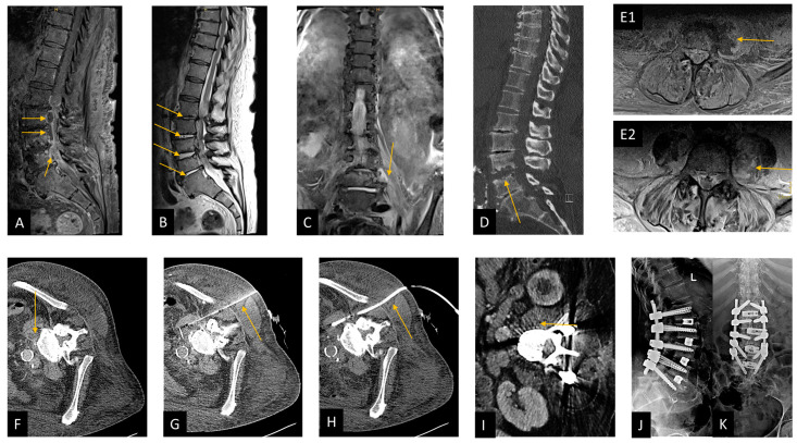
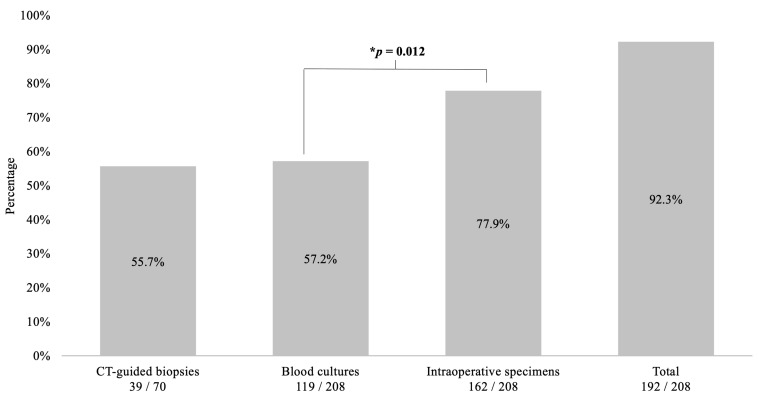
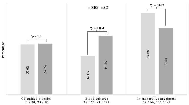
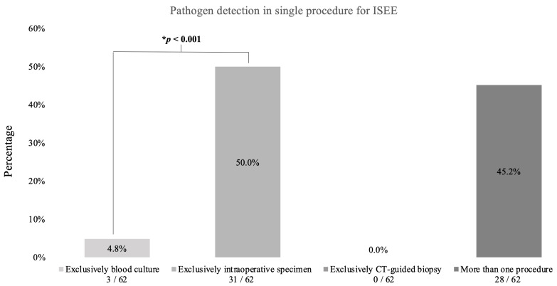
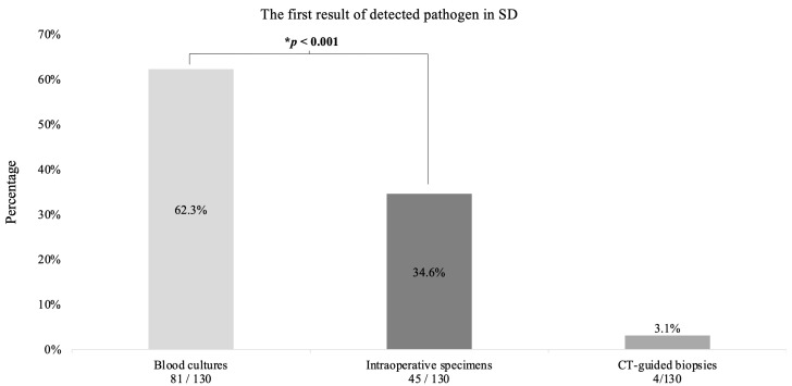
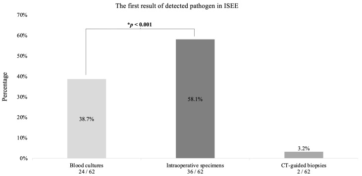
Results: we included 208 patients (68 [23-90] years, 34.6% females, 68% SD). Pathogens were identified in 192 cases (92.3%), including 187 (97.4%) pyogenic and five (2.6%) non-pyogenic infections, with Gram-positive bacteria accounting for 86.6% (162 cases) and Gram-negative for 13.4% (25 cases) of the pyogenic infections. The diagnostic sensitivity was highest for intraoperative specimens at 77.9% (162/208, p = 0.012) and lowest for blood cultures at 57.2% (119/208) and computed tomography (CT)-guided biopsies at 55.7% (39/70). Blood cultures displayed the highest sensitivity in SD patients (SD: 91/142, 64.1% vs. ISEE: 28/66, 42.4%, p = 0.004), while intraoperative specimens were the most sensitive procedure in ISEE (SD: 102/142, 71.8% vs. ISEE: 59/66, 89.4%, p = 0.007). The diagnostic sensitivity was lower in SD patients with ongoing empiric antibiotic therapy (EAT) than in patients treated postoperatively with targeted antibiotic therapy (TAT) (EAT: 77/89, 86.5% vs. TAT: 53/53, 100%, p = 0.004), whereas no effect was observed in patients with ISEE (EAT: 47/51, 92.2% vs. TAT: 15/15, 100%, p = 0.567).

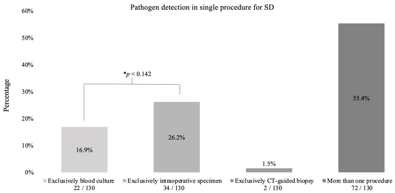
Conclusions: in our cohort, intraoperative specimens displayed the highest diagnostic sensitivity especially for ISEE, whereas blood cultures appear to be the most sensitive for SD. The sensitivity of these tests seems modifiable by preoperative EAT in patients with SD, but not in those with ISEE, underscoring the distinct differences between both pathologies.

Keywords: blood culture; computed tomography-guided biopsy; diagnostic sensitivity; intraoperative specimen; isolated spinal epidural empyema; spondylodiscitis.


Conflict of interest statement

The authors declare no conflict of interest.

Figures

Figure 1
Figure 1
Case presentation demonstrating CT-guided biopsy and surgical management in spondylodiscitis. This figure shows a patient from our cohort who suffered from spondylodiscitis with concomitant spinal epidural empyema and psoas abscess left; the 74-year-old patient was pretreated externally with calculated antibiotics (ceftriaxone, flucloxacillin, and metronidazole) and had paraparesis of the legs and sepsis; risk factors were a BMI greater than 35 kg/m and diabetes mellitus with chronic malum perforans pedis. Blood cultures were initially obtained from two different peripheral regions, followed immediately with microsurgical decompression with abscess evacuation and application of a suction-irrigation drainage system. CT-guided drainage of the psoas abscess was performed on the first postoperative day, whereas Staphelococcus aureus was detected only in blood culture and subsequently treated with flucloxacillin and rifampicin. Due to increasing bone destruction, transforaminal lumbar interbody fusion (TLIF) was performed at L1/L2, L2/L3, L3/4, and L4/L5 level. The patient was moved from the intensive care unit to the normal ward and mobilized at ward level. (A) Preoperative sagittal T1-weighted fat-saturated contrast-enhanced MRI image of the lumbar spine shows the epidural abscess in the spinal canal at L2–L4 level, marked with arrows. (B) Preoperative sagittal T2-weighted MRI image, arrows show spondylodiscitis at L1/L2, L2/L3, L3/L4, and L4/L5 level. (C) preoperative coronal T2-weighted short-tau inversion recovery (T2w-STIR) MRI image, arrow shows a psoas abscess on the left. (D) Preoperative sagittal reformated CT image, arrow shows bone destruction mainly at the level of L4/L5. (E1) Preoperative axial T1-weighted fat-saturated contrast-enhanced MRI image, arrow shows psoas abscess. (E2) Preoperative axial T2-weighted fat-saturated MRI image with arrow pointing to psoas abscess on the left. (FH): Illustration of CT-guided puncture of a left psoas abscess in three steps in an axial CT image. (F) Planning CT, (G) needle puncture, and (H) insertion of a suction-irrigation drain. Partially imaged central venous catheter in the iliac vein (G,H). (I) Postoperative axial CT image showing the regreening of the psoas abscess after draining the abscess. (J,K) A postoperative lateral (J) and anteroposterior (K) radiograph after performing TLIF spondylodesis from L1 to L5.
Figure 2
Figure 2
Sensitivity of blood culture, intraoperative specimen, and CT-guided biopsy. This figure shows the diagnostic sensitivity of the three procedures in combination and alone. The best results are obtained when all diagnostic procedures are used together, and the best individual result is achieved with intraoperative specimens, which are significantly more effective than blood cultures (77.9% vs. 57.2%, p = 0.012). * Binomial test. CT: computed tomography.
Figure 3
Figure 3
Diagnostic sensitivity in spondylodiscitis and isolated spinal epidural empyema. This figure shows the significant difference between both groups, especially in blood cultures (p = 0.004) and intraoperative specimens (p = 0.007). No difference was found between both groups concerning CT-guided biopsies. * Fisher exact test. ISEE: isolated spinal epidural empyema SD: spondylodiscitis, CT: computed tomography.
Figure 4
Figure 4
Pathogen detection in spondylodiscitis. This diagram shows in which procedures pathogens were detected in spondylodiscitis patients. There were 142 patients in total. Pathogens were detected in 130 patients. * Binomial test.
Figure 5
Figure 5
Pathogen detection in isolated spinal epidural empyema. This figure demonstrates the procedures used to detect the pathogens in epidural empyema patients. In total, there were 66 patients. Pathogens were detected in 62 patients. * Binomial test.
Figure 6
Figure 6
The first result of detected pathogen in spondylodiscitis. This diagram shows the first result of detected pathogen in Spondylodiszitis (SD) patients. * Binomial test.
Figure 7
Figure 7
The first result of detected pathogen in isolated spinal epidural empyema. This diagram reveals the first pathogen detection in isolated spinal epidural empyema (ISEE) patients. * Binomial test.

References

    1. Butler J.S., Shelly M.J., Timlin M., Powderly W.G., O’Byrne J.M. Nontuberculous pyogenic spinal infection in adults: A 12-year experience from a tertiary referral center. Spine. 2006;31:2695–2700. doi: 10.1097/01.brs.0000244662.78725.37. - DOI - PubMed
    1. Frangen T.M., Kalicke T., Gottwald M., Andereya S., Andress H.J., Russe O.J., Muller E.J., Muhr G., Schinkel C. Die operative Therapie der Spondylodiszitis. Unfallchirurg. 2006;109:743–753. doi: 10.1007/s00113-006-1084-7. - DOI - PubMed
    1. Tsiodras S., Falagas M.E. Clinical assessment and medical treatment of spine infections. Clin. Orthop. Relat. Res. 2006;444:38–50. doi: 10.1097/01.blo.0000203454.82264.cd. - DOI - PubMed
    1. Herren C., Jung N., Pishnamaz M., Breuninger M., Siewe J., Sobottke R. Spondylodiscitis: Diagnosis and Treatment Options. Dtsch. Arztebl. Int. 2017;114:875–882. doi: 10.3238/arztebl.2017.0875. - DOI - PMC - PubMed
    1. Zarghooni K., Rollinghoff M., Sobottke R., Eysel P. Treatment of spondylodiscitis. Int. Orthop. 2012;36:405–411. doi: 10.1007/s00264-011-1425-1. - DOI - PMC - PubMed

LinkOut - more resources