

A Systematic Review on Combined [18F]FDG and 68Ga-SSA PET/CT in Pulmonary Carcinoid
- PMID: 37297914
- PMCID: PMC10253705
- DOI: 10.3390/jcm12113719
A Systematic Review on Combined [18F]FDG and 68Ga-SSA PET/CT in Pulmonary Carcinoid
Abstract
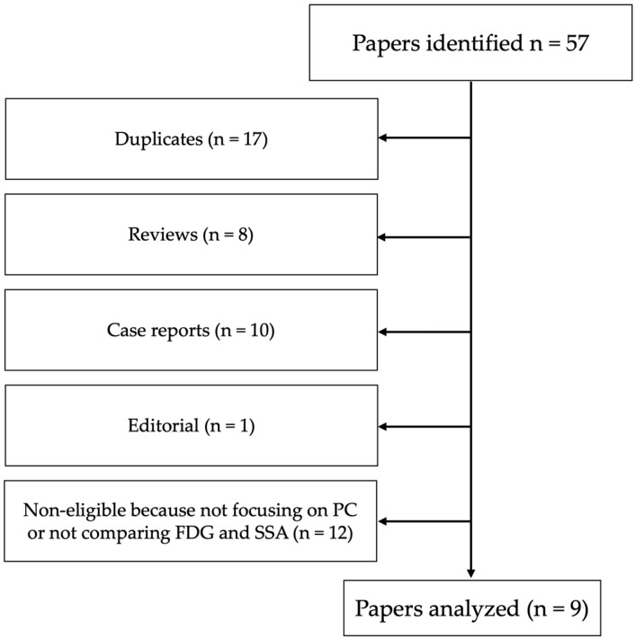
Pulmonary carcinoids (PCs) are part of a spectrum of well-differentiated neuroendocrine neoplasms (NENs) and are classified as typical carcinoid (TC) and atypical carcinoid (AC). TC differ from AC not only for its histopathological features but also for its "functional imaging pattern" and prognosis. ACs are more undifferentiated and characterized by higher aggressiveness. Positron emission tomography/computed tomography (PET/CT) with somatostatin analogs (SSA) labeled with Gallium-68 (68Ga-DOTA-TOC, 68Ga-DOTA-NOC, 68Ga-DOTA-TATE) has widely replaced conventional imaging with gamma camera using 111In- or 99mTc-labelled compounds and represents now the gold standard for diagnosis and management of NENs. In this setting, as already described for gastro-entero-pancreatic NENs, 18F-Fluorodeoxiglucose ([18F]FDG) in addition to 68Ga-SSA can play an important role in clinical practice, particularly for ACs that show a more aggressive behavior compared to TCs. The aim of this systematic review is to analyze all original studies collected from the PubMed and Scopus databases regarding PCs in which both 68Ga-SSA PET/CT and [18F]FDG PET/CT were performed in order to evaluate the clinical impact of each imaging modality. The following keywords were used for the research: "18F, 68Ga and (bronchial carcinoid or carcinoid lung)". A total of 57 papers were found, of which 17 were duplicates, 8 were reviews, 10 were case reports, and 1 was an editorial. Of the remaining 21 papers, 12 were ineligible because they did not focus on PC or did not compare 68Ga-SSA and [18F]FDG. We finally retrieved and analyzed nine papers (245 patients with TCs and 110 patients with ACs), and the results highlight the importance of the combined use of 68Ga-SSA and [18F]FDG PET/CT for the correct management of these neoplasms.
Keywords: 68Ga-SSA; [18F]FDG; atypical carcinoid; pulmonary carcinoid; typical carcinoid.
Conflict of interest statement
The authors declare no conflict of interest.
Figures
References
-
- Caplin M.E., Baudin E., Ferolla P., Filosso P., Garcia-Yuste M., Lim E., Oberg K., Pelosi G., Perren A., Rossi R.E., et al. Pulmonary neuroendocrine (carcinoid) tumors: European Neuroendocrine Tumor Society expert consensus and recommendations for best practice for typical and atypical pulmonary carcinoids. Ann Oncol. 2015;26:1604–1620. doi: 10.1093/annonc/mdv041. - DOI - PubMed
-
- Faggiano A., Ferolla P., Grimaldi F., Campana D., Manzoni M., Davì M.V., Bianchi A., Valcavi R., Papini E., Giuffrida D., et al. Natural history of gastro-entero-pancreatic and thoracic neuroendocrine tumors. Data from a large prospective and retrospective Italian epidemiological study: The NET management study. J. Endocrinol. Investig. 2012;35:817–823. - PubMed
-
- Thakur S., Florisson D., Telianidis S., Yaftian N., Lee J., Knight S., Barnett S., Seevanayagam S., Antippa P., Alam N., et al. Pulmonary carcinoid tumours: A multi-centre analysis of survival and predictors of outcome following sublobar, lobar, and extended pulmonary resections. Asian Cardiovasc. Thorac. Ann. 2021;29:532–540. doi: 10.1177/02184923211010090. - DOI - PubMed
Publication types
LinkOut - more resources
Full Text Sources
Research Materials
