. 2023 May 23;24(11):9118.
doi: 10.3390/ijms24119118.

Inducible and Conditional Activation of Adult Neurogenesis Rescues Cadmium-Induced Hippocampus-Dependent Memory Deficits in ApoE4-KI Mice

Affiliations

Inducible and Conditional Activation of Adult Neurogenesis Rescues Cadmium-Induced Hippocampus-Dependent Memory Deficits in ApoE4-KI Mice

Megumi T Matsushita et al. Int J Mol Sci. .

Abstract

The apolipoprotein E (ApoE) gene is a genetic risk factor for late-onset Alzheimer's disease, in which ε4 allele carriers have increased risk compared to the common ε3 carriers. Cadmium (Cd) is a toxic heavy metal and a potential neurotoxicant. We previously reported a gene-environment interaction (GxE) effect between ApoE4 and Cd that accelerates or increases the severity of the cognitive decline in ApoE4-knockin (ApoE4-KI) mice exposed to 0.6 mg/L CdCl2 through drinking water compared to control ApoE3-KI mice. However, the mechanisms underlying this GxE effect are not yet defined. Because Cd impairs adult neurogenesis, we investigated whether genetic and conditional stimulation of adult neurogenesis can functionally rescue Cd-induced cognitive impairment in ApoE4-KI mice. We crossed either ApoE4-KI or ApoE3-KI to an inducible Cre mouse strain, Nestin-CreERTM:caMEK5-eGFPloxP/loxP (designated as caMEK5), to generate ApoE4-KI:caMEK5 and ApoE3-KI:caMEK5. Tamoxifen administration in these mice genetically and conditionally induces the expression of caMEK5 in adult neural stem/progenitor cells, enabling the stimulation of adult neurogenesis in the brain. Male ApoE4-KI:caMEK5 and ApoE3-KI:caMEK5 mice were exposed to 0.6 mg/L CdCl2 throughout the experiment, and tamoxifen was administered once Cd-induced impairment in spatial working memory was consistently observed. Cd exposure impaired spatial working memory earlier in ApoE4-KI:caMEK5 than in ApoE3-KI:caMEK5 mice. In both strains, these deficits were rescued after tamoxifen treatment. Consistent with these behavioral findings, tamoxifen treatment enhanced adult neurogenesis by increasing the morphological complexity of adult-born immature neurons. These results provide evidence for a direct link between impaired spatial memory and adult neurogenesis in this GxE model.

Keywords: Alzheimer’s disease; adult neurogenesis; cadmium; gene–environment interaction; neurotoxicity.


Conflict of interest statement

The authors declare no conflict of interest.

Figures

Figure 1
Figure 1
Animal weights and water consumption of the behavioral cohort for ApoE3:caMEK5 and ApoE4:caMEK5 mice. (A) Temporary body weight loss was observed in tamoxifen-treated ApoE3-KI:caMEK5 and ApoE4-KI:caMEK5 mice. (B) Tamoxifen treatment did not affect water consumption in ApoE3-KI:caMEK5 or ApoE4-KI:caMEK5 mice. Bars (red) indicate the time of tamoxifen treatment. Data are presented as mean ± SEM.
Figure 2
Figure 2
Effects of Cd exposure on hippocampus-dependent short-term spatial memory in the novel object location (NOL) test. (A) Schematic of NOL test. During training, two identical objects were placed in two adjacent locations A and B. The duration of time each mouse spent exploring identical objects in the old location A and novel location C was quantified in the test session. (B) ApoE4-KI and ApoE3-KI mice were unable to distinguish between the old and new locations starting at 16 and 28 weeks of Cd exposure, respectively. However, this impairment was reversed after tamoxifen, but not vehicle (veh) control treatment. Cd treatment lasted throughout the experiment (gray box); time points after vehicle/tamoxifen treatment are boxed in light purple. Each NOL test time point is indicated with an arrowhead. The observed NOL impairments are indicated by red arrowheads while restored memory tests are indicated by purple arrowheads. Representative data for % exploration time were shown for select time points (lines linked to the arrowheads). Data are presented as mean ± SEM. Welch’s two sample t-test: * p ≤ 0.05, ** p ≤ 0.01, *** p ≤ 0.001, **** p ≤ 0.0001.
Figure 3
Figure 3
Cd concentrations in mouse blood and brain hemisphere at the end of the experiment in the behavioral cohort after 54 weeks (ApoE3-KI:caMEK5) or 49 weeks of exposure (ApoE4-KI:caMEK5). There were no significant differences between vehicle- and tamoxifen-treated groups in (A) blood Cd of ApoE3-KI, (B) blood Cd of ApoE4-KI, or (C) brain Cd of ApoE3-KI. (D) Tamoxifen-treated ApoE4-KI mice had statistically significant higher brain Cd levels compared to vehicle-treated ApoE4-KI mice. Data are presented as mean ± SEM. Welch’s two sample t-test: * p ≤ 0.05.
Figure 4
Figure 4
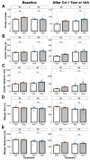
Locomotor activity of ApoE3-KI and ApoE4-KI mice (n = 12–15/group) was measured in the open field test at baseline and after Cd + Veh or Tam treatment. (A) number of moves. (B) total moving time. (C) total moving distance. Data are presented as mean ± SEM. Welch’s two sample t-test: * p ≤ 0.05.
Figure 5
Figure 5
Anxiety behavior of ApoE3-KI and ApoE4-KI mice (n = 12–15/group) was measured in the open field test at baseline and after Cd + Veh or Tam treatment. (A) number of entries to the center, (B) center time, (C) center distance, (D) margin time, or (E) margin distance. Data are presented as mean ± SEM. Welch’s two-sample t-test: * p ≤ 0.05.
Figure 6
Figure 6
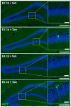
Representative images of TSA signal amplified caMEK5-eGFP signal. Full DG field images were taken on a 20× objective and stitched in LAS X; scale bar: 200 µm. Inlay: Zoomed-in images from ROI; scale bar: 30 µm.
Figure 7
Figure 7
Quantification of adult-born neurons that differentiated into mature neurons in the DG of the hippocampus. (A) Representative images of BrdU+ (red) and NeuN+ (green) staining. Scale bar, 30 µm. Quantification of (B) total number of BrdU+ cells, (C) total number of BrdU+ and NeuN+ cells, and (D) ratio of BrdU+ and NeuN+ cells to BrdU+ cells for tamoxifen- or vehicle-treated ApoE3-KI:caMEK5 (n = 4–5) and ApoE4-KI:caMEK5 (n = 6–7) mice. Data are presented as mean ± SEM.
Figure 8
Figure 8
Quantification of adult-born immature neurons in the DG of the hippocampus. (A) Representative images of BrdU+ (red) and DCX+ (green) staining. Scale bar, 30 µm. Quantification of (B) total number of BrdU+ cells, (C) total number of BrdU+ and DCX+ cells, and (D) ratio of BrdU+ and DCX+ cells to BrdU+ cells for tamoxifen- or vehicle-treated ApoE3-KI:caMEK5 (n = 4–5) and ApoE4-KI:caMEK5 (n = 6–7) mice. Data are presented as mean ± SEM.
Figure 9
Figure 9
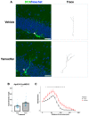
Dendritic branching of DCX+ cells in the DG of ApoE3-KI:caMEK5 mice. (A) Representative images (arrowhead) of cell tracing of immature neurons with Hoechst (blue) and DCX (green) staining in the DG. Scale bar, 30 µm. (B) Tamoxifen-treated animals have significantly increased total dendritic length compared to vehicle-treated animals. Welch’s two sample t-test: *** p ≤ 0.001. (C) Tamoxifen-treated animals have significantly increased numbers of intersections compared to vehicle-treated animals. Pairwise comparisons at each experimental week were based on estimates from mixed-effects linear regression, with Tukey HSD correction: * p ≤ 0.05, ** p ≤ 0.01, *** p ≤ 0.001. n = 3–4 animals per group, 39–44 cells per group.
Figure 10
Figure 10
Dendritic branching of DCX+ cells in the DG of ApoE4-KI:caMEK5 mice. (A) Representative images (arrowhead) of cell tracing of immature neurons with Hoechst (blue) and DCX (green) staining in the DG. Scale bar, 30 µm. (B) Tamoxifen-treated animals have significantly increased total dendritic length compared to vehicle-treated animals. Welch’s two sample t-test: **** p ≤ 0.0001. (C) Tamoxifen-treated animals have significantly increased numbers of intersections compared to vehicle-treated animals. Pairwise comparisons at each experimental week were based on estimates from mixed-effects linear regression, with Tukey HSD correction: * p ≤ 0.05, ** p ≤ 0.01, *** p ≤ 0.001. N = 4 animals per group, 45–52 cells per group.
Figure 11
Figure 11
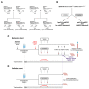
Study design for the caMEK5 rescue experiment. (A) Breeding scheme. (B) The schematic diagram for generation of transgenic mice through tamoxifen treatment. Tamoxifen-treated animals express constitutively active MEK5 (caMEK5) in adult neurogenic regions (under the Nestin-CreERTM promoter) with ApoE4-KI background (ApoE4-KI:caMEK5); control animals have the same genetic background but do not express caMEK5 (ApoE4-KI:control). The same strategy was used to generate ApoE3-KI:caMEK5 and control mice. (C) Study design for the caMEK5 rescue experiment within each genotype in the behavior cohorts. Prior to Cd treatment, baseline behavior was assessed with the open field and novel object location (NOL) tests. All animals received Cd through drinking water (0.6 mg/L CdCl2) from 8–10 weeks of age until the time of euthanasia. Cognitive function was probed every other week with a NOL test until a deficit was observed, at which time, NOL was performed weekly. Once memory deficits were confirmed by the NOL test at three consecutive time points, animals were given tamoxifen (n = 12–15/group) treatment to induce caMEK5 expression. The control group received a vehicle that was used to dissolve tamoxifen and thus should not express caMEK5. Four weeks after the last dose of tamoxifen or vehicle administration, the NOL test was performed to assess cognitive function after tamoxifen or vehicle treatment. The open field test was performed after the functional rescue was confirmed in at least three consecutive NOL tests. At the end of the experiment, blood and brain tissues were collected for Cd analysis. (D) Study design for the caMEK5 rescue experiment within each genotype in the cellular cohorts. Tissue collection in the cellular cohort was based on the last date of the three consecutive time points of NOL rescue confirmation in the behavior cohort.

Similar articles

Cited by

References

    1. Alzheimer’s Association 2022 Alzheimer’s Disease Facts and Figures. Alzheimers Dement. 2022;18:700–789. doi: 10.1002/alz.12638. - DOI - PubMed
    1. Liu C.-C., Kanekiyo T., Xu H., Bu G. Apolipoprotein E and Alzheimer Disease: Risk, Mechanisms and Therapy. Nat. Rev. Neurol. 2013;9:106–118. doi: 10.1038/nrneurol.2012.263. - DOI - PMC - PubMed
    1. Bekris L.M., Yu C.-E., Bird T.D., Tsuang D.W. Genetics of Alzheimer Disease. J. Geriatr. Psychiatry Neurol. 2010;23:213–227. doi: 10.1177/0891988710383571. - DOI - PMC - PubMed
    1. Bakulski K.M., Seo Y.A., Hickman R.C., Brandt D., Vadari H.S., Hu H., Park S.K. Heavy Metals Exposure and Alzheimer’s Disease and Related Dementias. J. Alzheimers Dis. 2020;76:1215–1242. doi: 10.3233/JAD-200282. - DOI - PMC - PubMed
    1. Migliore L., Coppedè F. Gene–Environment Interactions in Alzheimer Disease: The Emerging Role of Epigenetics. Nat. Rev. Neurol. 2022;18:643–660. doi: 10.1038/s41582-022-00714-w. - DOI - PubMed