. 2023 May 24;24(11):9216.
doi: 10.3390/ijms24119216.

Procoagulant Activity of Umbilical Cord-Derived Mesenchymal Stromal Cells' Extracellular Vesicles (MSC-EVs)

Affiliations

Procoagulant Activity of Umbilical Cord-Derived Mesenchymal Stromal Cells' Extracellular Vesicles (MSC-EVs)

Adrienne Wright et al. Int J Mol Sci. .

Abstract

Many cell types, including cancer cells, release tissue factor (TF)-exposing extracellular vesicles (EVs). It is unknown whether MSC-EVs pose a thromboembolism risk due to TF expression. Knowing that MSCs express TF and are procoagulant, we hypothesize that MSC-EVs also might. Here, we examined the expression of TF and the procoagulant activity of MSC-EVs and the impact of EV isolation methods and cell culture expansion on EV yield, characterization, and potential risk using a design of experiments methodology. MSC-EVs were found to express TF and have procoagulant activity. Thus, when MSC-derived EVs are employed as a therapeutic agent, one might consider TF, procoagulant activity, and thromboembolism risk and take steps to prevent them.

Keywords: canine; clinical safety; exosome; hemocompatibility; human; mesenchymal stromal cells; tissue factor.


Conflict of interest statement

The authors declare no conflict of interest. The funders had no role in the design of the study; in the collection, analyses, or interpretation of data; in the writing of the manuscript, or in the decision to publish the results.

Figures

Figure 1
Figure 1
Protein concentration of EV samples. (AC) The effect of passage on protein content was not obvious. However, a frank difference was noted between the protein contents of size-exclusion chromatography-isolated EVs (SEC, white bars) and ultracentrifugation-isolated EVs (UC, gray bars). (D,E) When protein content was plotted vs. cumulative population doublings (CPD), the trend lines suggested that protein content in EVs tended to increase with longer passage times. This trend was more apparent in SEC-isolated EVs. (F) When we studied whether protein content was affected by population doubling time, no big impact of population doubling time was noted. (GI) When the particles per ug protein were calculated, UC and SEC isolated EVs had fewer particles per ug protein in late passages. The asterisk indicates p < 0.05.
Figure 2
Figure 2
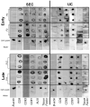
Extracellular vesicles (EVs) isolated from canine umbilical cord-derived mesenchymal stromal cells (MSCs) express tissue factor (TF) over passages but differ in terms of the expression of CD63 between early and late passages. Both early (P2 and P3 shown) and late passage (P11 and P12 shown) EVs expressed TF regardless of the EV isolation method (size-exclusion chromatography, SEC, or ultracentrifugation, UC). The expression of clusters of differentiation (CD) 9, CD63, CD81; ALIX; TF; protein loading control, ß-actin, via negative control (water), whole-MSC cell lysate (positive control) in both early (top) and late (bottom) passages, as well as EVs isolated via SEC (left) and UC (right). EVs express CD9, CD81, and ALIX regardless of passaging or isolation. However, irrespective of the isolation method, CD63 expression was not observed in late passage EVs.
Figure 3
Figure 3
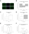
Procoagulant activity of MSCs and EVs. (A) Tissue factor (TF) was expressed by human and canine umbilical cord-derived mesenchymal stromal cells. Note that the staining intensity of human MSCs (bottom) appears to be higher than that of canine MSCs. Calibration bar = 400 µM. (B) The procoagulant activity (FXa generation) of human and canine extracellular vesicles (EVs) isolated from umbilical cord mesenchymal stromal cells (MSCs). No significant differences in procoagulant activity levels were found between human MSCs (positive control, left bar) and canine (middle) or human EVs. Data from three human MSC lines from passages 4 to 6 and three human and canine EV samples. (C) Procoagulant activity (ng/mL of FXa generated) of EVs isolated from canine umbilical cord MSCs via cell passaging: early (P2–P5) vs. late (P9–P12) passage. No statistical differences were found, but the trend was for late-passage EVs to have higher levels of procoagulant activity. (D) No significant differences were found between the isolation method used: size exclusion chromatography- (SEC, white bars) vs. ultracentrifugation (UC, gray bars)-based EV isolation. There was a trend for EVs isolated via UC to have a higher procoagulant activity level than that of SEC. Data from EVs were derived from six canine MSC lines; there were three in each isolation method. (E) Canine EVs had significantly higher procoagulant activity levels than the negative control did (FX + FVIIa). However, when they were incubated with polyclonal anti-tissue factor antibody, FX-a generation was not inhibited in K9 EVs. (F) Human EVs displayed significantly more procoagulant activity than the negative control did (FX + FVIIa). The procoagulant activity of human EVs was significantly inhibited by an anti-TF antibody (clone HTF-1 was previously shown to inhibit the function of TF). In contrast, and similar to canine EVs, procoagulant activity was not inhibited by the polyclonal TF antibody (far right). Data are presented as the average of three biological replicates ± standard deviation. The asterisk indicates p < 0.05.
Figure 4
Figure 4
Experimental design and schematic. A randomized, 8 × 2 factorial is shown. (A). Design of experiments protocol parsed into two factors. Factor A (Cell Passage) had eight levels: P2, P3, P4, P5, P9, P10, P11, and P12. Factor B (EV isolation method) had two levels: ultracentrifugation (UC) and size-exclusion chromatography (SEC). Six canine umbilical cord-derived mesenchymal stromal cell lines (CUC) selected from a cell bank were used and randomly assigned to levels of Factor B. (B). Experimental schematic: Canine umbilical cord-derived mesenchymal stromal cells (MSCs) were thawed at passage 1 (P1), plated at a density on gelatin-coated T-150 flasks, and cultured as previously described [58]. Once plates reached 80–90% confluence, MSCs were passaged and plated into two T-150 flasks. Flask (1) was used for the production of conditioned medium (CM) (arrow going to the left side): cell culture medium of MSCs at 60–70% confluence was removed and replaced with DMEM for 24 h. This medium was considered to be “conditioned” and was collected in a 50 mL centrifuge tube. The CM was stored at −80 °C until isolation via size-exclusion chromatography (SEC) or ultracentrifugation (UC) [59]. Flask (2) was used to maintain the MSC line in culture (arrow pointing to the right side). In Factor A, passages 2–5 were defined as “early passage”, and passages 9–12 were described as “late passage”.

References

    1. Pittenger M.F., Discher D.E., Peault B.M., Phinney D.G., Hare J.M., Caplan A.I. Mesenchymal stem cell perspective: Cell biology to clinical progress. NPJ Regen. Med. 2019;4:22. doi: 10.1038/s41536-019-0083-6. - DOI - PMC - PubMed
    1. Levy O., Kuai R., Siren E.M.J., Bhere D., Milton Y., Nissar N., De Biasio M., Heinelt M., Reeve B., Abdi R., et al. Shattering barriers toward clinically meaningful MSC therapies. Sci. Adv. 2020;6:eaba6884. doi: 10.1126/sciadv.aba6884. - DOI - PMC - PubMed
    1. Galipeau J., Sensebe L. Mesenchymal Stromal Cells: Clinical Challenges and Therapeutic Opportunities. Cell Stem Cell. 2018;22:824–833. doi: 10.1016/j.stem.2018.05.004. - DOI - PMC - PubMed
    1. Durand N., Mallea J., Zubair A.C. Insights into the use of mesenchymal stem cells in COVID-19 mediated acute respiratory failure. NPJ Regen. Med. 2020;5:17. doi: 10.1038/s41536-020-00105-z. - DOI - PMC - PubMed
    1. Djouad F., Bouffi C., Ghannam S., Noel D., Jorgensen C. Mesenchymal stem cells: Innovative therapeutic tools for rheumatic diseases. Nat. Rev. Rheumatol. 2009;5:392–399. doi: 10.1038/nrrheum.2009.104. - DOI - PubMed

MeSH terms

LinkOut - more resources