. 2023 May 25;24(11):9221.
doi: 10.3390/ijms24119221.

The Effect of Statins on Male Reproductive Parameters: A Mechanism Involving Dysregulation of Gonadal Hormone Receptors and TRPV1

Affiliations

The Effect of Statins on Male Reproductive Parameters: A Mechanism Involving Dysregulation of Gonadal Hormone Receptors and TRPV1

Temidayo S Omolaoye et al. Int J Mol Sci. .

Abstract

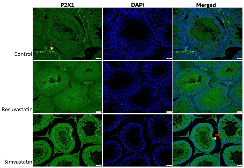
Statins have been shown to cause diverse male reproductive function impairment, and in some cases, orchialgia. Therefore, the current study investigated the possible mechanisms through which statins may alter male reproductive parameters. Thirty adult male Wistar rats (200-250 g) were divided into three groups. The animals were orally administered rosuvastatin (50 mg/kg), simvastatin (50 mg/kg), or 0.5% carboxy methyl cellulose (control), for a 30-day period. Spermatozoa were retrieved from the caudal epididymis for sperm analysis. The testis was used for all biochemical assays and immunofluorescent localization of biomarkers of interest. Rosuvastatin-treated animals presented with a significant decrease in sperm concentration when compared to both the control and simvastatin groups (p < 0.005). While no significant difference was observed between the simvastatin and the control group. The Sertoli cells, Leydig cells and whole testicular tissue homogenate expressed transcripts of solute carrier organic anion transporters (SLCO1B1 and SLCO1B3). There was a significant decrease in the testicular protein expression of the luteinizing hormone receptor, follicle stimulating hormone receptor, and transient receptor potential vanilloid 1 in the rosuvastatin and simvastatin-treated animals compared to the control. The expression of SLCO1B1, SLCO1B2, and SLCO1B3 in the different spermatogenic cells portray that un-bio transformed statin can be transported into the testicular microenvironment, which can subsequently alter the regulation of the gonadal hormone receptors, dysregulate pain-inflammatory biomarkers, and consequently impair sperm concentration.

Keywords: inflammation; male infertility; rosuvastatin; sex hormone receptors; simvastatin; testicular pain.


Conflict of interest statement

The authors declare no conflict of interest.

Figures

Figure 1
Figure 1
Effects of rosuvastatin and simvastatin on sperm parameters. (A) Sperm concentration, n = 10; (B) Percentage of morphologically normal spermatozoa, n = 10. M/mL—million/mL.
Figure 2
Figure 2
Relative mRNA expression of gonadal hormone receptors (LHR, FSHR) and solute carrier transporters (SLCO1B1, SLCO1B2 and SLCO1B3) in rat Leydig and Sertoli cells. The figure confirms that two of the most important cells (Sertoli and Leydi cells) involved in spermatogenesis maintenance and steroidogenesis, respectively, express LHR, FSHR, and different solute carrier organic anion transporters. These findings enunciate the possibility of their activation and involvement in diverse conditions upon appropriate stimulation. It is important to note that these are baseline values showing the presence of these genes in these cells. Values are representative of a minimum of three biological replicates.
Figure 3
Figure 3
The effect of rosuvastatin and simvastatin on testis mRNA expression of SLCO1B1, SLCO1B2, and SLCO1B3. (A) Relative mRNA of SLCO1B2 in the testes. (B) Relative mRNA of SLCO1B1 in the testes. (C) Relative mRNA of SLCO1B3 in the testes. SLCO1B1—solute carrier organic anion transporter superfamily 1B1; SLCO1B2—solute carrier organic anion transporter superfamily 1B2; SLCO1B3—solute carrier organic anion transporter superfamily 1B3. Values are representative of a minimum of three biological replicates (n = 3–6).
Figure 4
Figure 4
Effect of rosuvastatin and simvastatin on testicular LH and FSH receptors. (A) Western blot images of LHR and FSHR. (B) Normalized expression of LHR. (C) Normalized expression of FSHR. The first lane in the immunoblots is the ladder (molecular weight), while the remaining lanes represent samples from control (n = 3), simvastatin (n = 3), and rosuvastatin (n = 3). Overall, the number of samples reported per group on the graphs include a minimum of six. LHR = Luteinizing hormone receptor; FSHR = follicle stimulating hormone receptor.
Figure 5
Figure 5
The effect of rosuvastatin and simvastatin on the testicular mRNA expression of pain receptors. (A) Relative testicular mRNA expression of TRPV1. (B) Relative testicular mRNA expression of P2X1. (C) Relative testicular mRNA expression of P2X3. TRPV1—transient receptor potential vanilloid 1 gene; P2X1—purinergic receptor 1 gene; P2X3—purinergic receptor 3 gene. Values are representative of a minimum of four biological replicates (n = 4–6).
Figure 6
Figure 6
The effect of rosuvastatin and simvastatin on the testicular protein expression of pain receptors. (A) Western Blot images of TRPV1, P2X2 and P2X3. (B) Testicular protein expression of TRPV1. (C) Testicular protein expression of P2X1. (D) Testicular protein expression of P2X3. The first lane in the immunoblots is the ladder, while the remaining lanes represent samples from control (n = 3), simvastatin (n = 3), and rosuvastatin (n = 3). Overall, the number of samples reported per group on the graphs include a minimum of six biological replicates.
Figure 7
Figure 7
The effect of rosuvastatin and simvastatin on the expression of inflammatory cytokines. (A) immunoblot of GP130. (B) The protein expression of GP130 in the testes. (C) The mRNA expression of IL-6 in the testes. (D) The mRNA expression of testicular IL-17B. (E) The mRNA expression of testicular IL-17A. The first lane in the immunoblots is the ladder, while the remaining lanes represent samples from control (n = 3), simvastatin (n = 3), and rosuvastatin (n = 3). Overall, the number of samples reported per group in the graphs include a minimum of six. For the transcript expression, values are representative of four to six biological replicates.
Figure 8
Figure 8
The expression of LHR in the testes. As shown in the figure, LHR is mostly localized in the testicular interstitial, as the different cells in this environment have positive immunoreactivity to LHR. Fluorescence green = immunoreactivity of the protein; DAPI = a counter stain (stains nucleus blue); BV = Blood vessel; CC = connective cell or fibroblast; LC = Leydig cell. Scale bar—50 µm, Magnification—400×.
Figure 9
Figure 9
The expression of FSHR in the testes. The Sertoli and interstitial cells express FSHR and there is more positive immunoreactivity to FSHR in the control group compared to both rosuvastatin and simvastatin. Fluorescence green = immunoreactivity of the protein; DAPI = a counter stain (stains nucleus blue); SC = Sertoli cell; IC = Interstitial cells. Scale bar—50 µm, Magnification—400×.
Figure 10
Figure 10
The expression of TRPV1 in the testes. The interstitial cells and primary spermatocytes are immunoreactive to TRPV1. Fluorescence green = immunoreactivity of the protein; DAPI = a counter stain (stains nucleus blue); IC = Interstitial or stroma cells; PS = Primary spermatocyte. Scale bar—50 µm, Magnification—400×.
Figure 11
Figure 11
The expression of P2X1 in the testes. P2X1 is expressed in primary spermatocytes and the interstitial cells of the testes. No observable differences are seen in fluorescent intensity between the three groups. Fluorescence green = immunoreactivity of the protein; DAPI = a counter stain (stains nucleus blue); IC = Interstitial cells, PS = Primary spermatocyte. Scale bar—50 µm, Magnification—400×.
Figure 12
Figure 12
The expression of P2X3 in the testes. P2X3 is expressed in the spermatogonia and primary spermatocytes of the testes. Spermatogonia and primary spermatocytes of both rosuvastatin and simvastatin groups displayed more positive immunoreactivity to P2X3 compared to the control group. Fluorescence green = immunoreactivity of the protein; DAPI = a counter stain (stains nucleus blue); PS = Primary spermatocyte, SG = Spermatogonia, Scale bar—50 µm, Magnification—400×.
Figure 13
Figure 13
The expression of GP130 in the testes. Myoid cells, Leydig cells and blood vessels show immunoreactivity to GP130. Fluorescence green = immunoreactivity of the protein; DAPI = a counter stain (stains nucleus blue); MC = Myoid cells, LC = Leydig cell, BV = Blood vessel, Scale bar—50 µm, Magnification—400×.

References

    1. Navarese E.P., Buffon A., Andreotti F., Kozinski M., Welton N., Fabiszak T., Caputo S., Grzesk G., Kubica A., Swiatkiewicz I., et al. Meta-analysis of impact of different types and doses of statins on new-onset diabetes mellitus. Am. J. Cardiol. 2013;111:1123–1130. doi: 10.1016/j.amjcard.2012.12.037. - DOI - PubMed
    1. Leite G.A.A., de lima Rosa J., Sanabria M., Cavariani M.M., Franci J.A.A., Pinheiro P.F.F., Kempinas W.D.G. Delayed reproductive development in pubertal male rats exposed to the hypolipemiant agent rosuvastatin since prepuberty. Reprod. Toxicol. 2014;44:93–103. doi: 10.1016/j.reprotox.2014.01.004. - DOI - PubMed
    1. Leite G.A.A., de Barros J.W.F., de Martins A.C., Anselmo-Franci J.A., Barbosa F., Kempinas W.D.G. Ascorbic acid supplementation ameliorates testicular hormonal signaling, sperm production and oxidative stress in male rats exposed to rosuvastatin during pre-puberty. J. Appl. Toxicol. 2019;39:305–321. doi: 10.1002/jat.3720. - DOI - PubMed
    1. Leite G.A.A., Figueiredo T.M., Pacheco T.L., Sanabria M., e Silva P.V., Fernandes F.H., Kempinas W.D.G. Vitamin C partially prevents reproductive damage in adult male rats exposed to rosuvastatin during prepuberty. Food Chem. Toxicol. 2017;109:272–283. doi: 10.1016/j.fct.2017.09.003. - DOI - PubMed
    1. E Silva P.V., Borges C.D.S., Rosa J.D.L., Pacheco T.L., Figueiredo T.M., Leite G.A.A., Guerra M.T., Anselmo-Franci J.A., Klinefelter G.R., Kempinas W.D.G. Effects of isolated or combined exposure to sibutramine and rosuvastatin on reproductive parameters of adult male rats. J. Appl. Toxicol. 2020;40:947–964. doi: 10.1002/jat.3955. - DOI - PubMed