. 2023 May 27;24(11):9359.
doi: 10.3390/ijms24119359.

A Unique Mode of Coenzyme A Binding to the Nucleotide Binding Pocket of Human Metastasis Suppressor NME1

Affiliations

A Unique Mode of Coenzyme A Binding to the Nucleotide Binding Pocket of Human Metastasis Suppressor NME1

Maria-Armineh Tossounian et al. Int J Mol Sci. .

Abstract

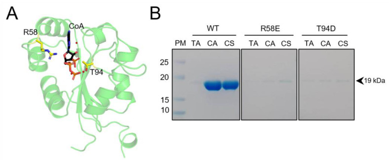
Coenzyme A (CoA) is a key cellular metabolite which participates in diverse metabolic pathways, regulation of gene expression and the antioxidant defense mechanism. Human NME1 (hNME1), which is a moonlighting protein, was identified as a major CoA-binding protein. Biochemical studies showed that hNME1 is regulated by CoA through both covalent and non-covalent binding, which leads to a decrease in the hNME1 nucleoside diphosphate kinase (NDPK) activity. In this study, we expanded the knowledge on previous findings by focusing on the non-covalent mode of CoA binding to the hNME1. With X-ray crystallography, we solved the CoA bound structure of hNME1 (hNME1-CoA) and determined the stabilization interactions CoA forms within the nucleotide-binding site of hNME1. A hydrophobic patch stabilizing the CoA adenine ring, while salt bridges and hydrogen bonds stabilizing the phosphate groups of CoA were observed. With molecular dynamics studies, we extended our structural analysis by characterizing the hNME1-CoA structure and elucidating possible orientations of the pantetheine tail, which is absent in the X-ray structure due to its flexibility. Crystallographic studies suggested the involvement of arginine 58 and threonine 94 in mediating specific interactions with CoA. Site-directed mutagenesis and CoA-based affinity purifications showed that arginine 58 mutation to glutamate (R58E) and threonine 94 mutation to aspartate (T94D) prevent hNME1 from binding to CoA. Overall, our results reveal a unique mode by which hNME1 binds CoA, which differs significantly from that of ADP binding: the α- and β-phosphates of CoA are oriented away from the nucleotide-binding site, while 3'-phosphate faces catalytic histidine 118 (H118). The interactions formed by the CoA adenine ring and phosphate groups contribute to the specific mode of CoA binding to hNME1.

Keywords: CoAlation; NDPK-A structure; NM23-H1; NME1; X-ray crystallography; coenzyme A; metastasis suppressor; molecular dynamics; nucleotide binding; protein-metabolite regulation.


Conflict of interest statement

The authors declare no conflict of interest.

Figures

Figure 1
Figure 1
Human NME1 moonlighting functions and redox regulation. (A) The different reported functions of human NME1 are shown. The structure of NME1 (PDB: 2HVD) is presented in a green Cartoon, the cysteine residues (C4, C109 and C145) in yellow Sticks, and the catalytic histidine (H118) in green Sticks. The Kpn loop is indicated in orange, and the nucleotide-binding (NB) site is shown in black dashed circle. The different types of redox modifications observed on NME1 are shown and include sulfonylation, CoAlation, glutathionylation and intramolecular disulfide bond formation. (B) The CoA structure is composed of a 3′-phosphorylated ADP moiety (black dashed box) and a long pantetheine tail with a reactive thiol group at its terminal (orange dashed box). CoA structure was generated using ChemDraw.
Figure 2
Figure 2
Purification and co-crystallization of hNME1. (A) The hNME1 SEC chromatogram is shown, where the blue line represents the absorbance at 280 nm. The purity of the SEC peak fractions was assessed by SDS-PAGE. The insert shows the reducing SDS-PAGE gel with purified hNME1 bands around 19 kDa. The molecular weight (kDa) of the protein markers is indicated. (B) hNME1-CoA crystals are shown.
Figure 3
Figure 3
CoA binds non-covalently to the hNME1 nucleotide-binding site. (A) The hexameric structure (dimer of trimers) of hNME1-CoA is presented, where the top three monomers (a (green Cartoon), b (yellow Cartoon) and c (orange Cartoon)) forming a trimer are shown in a black dashed rectangle. The bottom three monomers of the hexamer are indicated as a’ (dark green Cartoon), b’ (light orange Cartoon) and c’ (bright orange Cartoon). Each monomer contains a CoA molecule. (B) Overlay of the three hNME1-CoA monomers (a, b and c). The α-, β- and 3′-phosphates of CoA are indicated. The β-phosphate of CoA-b has a different orientation than the β-phosphates of CoA-a/c. (C) Surface view of hNME1-CoA monomer a. The insert shows the 2Fo-Fc electron density map surrounding the adenosine part of CoA molecule at 1.2σ contour level. The density surrounding the pantetheine tail is not observed. CoA is shown in black or green Sticks and colored by element, where the nitrogen, oxygen, and phosphorous atoms are in blue, red, and orange colors, respectively.
Figure 4
Figure 4
CoA stabilization interactions within the hNME1 nucleotide-binding site. The stabilization interactions of CoA within the NB site of hNME1-CoA monomer a (green Cartoon) are shown (AC). The residues that form hydrophobic interactions are colored green, and the residues that form salt bridges or H-bonds (in presence or absence of a water molecule (W)—red sphere) are shown in yellow. (A) Interactions between the rings of the ADP moiety of CoA and the hydrophobic patch of the NB site are shown. Interactions between the adenine ring and K12 through a water molecule are shown in black dotted line. H-bond between the hydroxyl group of the ribose ring and a water molecule (W) is shown in orange dotted line. H-bond interactions between the hNME1 NB site residues and (B) the 3′-phosphate and (C) α-/β-phosphates of CoA are shown in black dotted lines. CoA is shown in Sticks and colored by element, where the carbon, nitrogen, oxygen, and phosphorous atoms are represented in black, blue, red, and orange colors, respectively.
Figure 5
Figure 5
CoA binding induces a local shift of loop56–60 and the hydrophobic patch towards the NB site. Structural overlay of hNME1-apo (cyan Cartoon; PDB: 1JXV), hNME1-CoA (green Cartoon) and hNME1-ADP (wheat Cartoon; PDB: 2HVD) (AC) is shown. (A,B) Residues on the loop56–60 and near the α-helices located directly before and after the loop shift closer to the center of the NB site to stabilize CoA and ADP. The shift is indicated with a black arrow. (C) CoA α- and β-phosphates are oriented away from the catalytic center compared to the phosphates of ADP (black arrow). (DF) Structural overlay of hNME1-apo (cyan Cartoon; PDB: 1JXV), hNME1-CoA (green Cartoon) and hNME2-mCoA (pink Cartoon; PDB: 7KPF) is shown. (D,E) Residues on the loop56–60 and near the α-helices located directly before and after the loop shift closer to the center of the NB site to stabilize CoA and mCoA (black arrow). (F) mCoA (pink) α- and β-phosphates are also oriented away from the catalytic center, similar to CoA-a (black) and CoA-b (yellow). The CoA-a, CoA-b, ADP and mCoA are colored by element, where carbon atoms are in black, yellow, wheat and pink, respectively. Nitrogen, oxygen, and phosphorous atoms are represented in blue, red, and orange colors, respectively.
Figure 6
Figure 6
CoA and myristoyl-CoA α- and β-phosphate groups are oriented away from the catalytic pocket. (A) Overlay of the nucleotide containing structures of nucleotide kinases. The α- and β-phosphate groups of the nucleotides are oriented towards the catalytic H118. (B) CoA and (C) myristoyl-CoA (mCoA) α- and β-phosphate groups are oriented away from the catalytic pocket (black arrow). The nucleotide structures are shown in “Lines”, while CoA and mCoA are shown in Sticks. The ribose units of CoA and mCoA are shifted (black dotted arrow) compared to the other bound nucleotides. The nucleotides, CoA and mCoA are colored by element, where carbon atoms are in gray, green and black, respectively. Nitrogen, oxygen, and phosphorous atoms are represented in blue, red, and orange colors, respectively.
Figure 7
Figure 7
Molecular dynamics-based characterization of the hNME1-CoA interaction in aqueous solution. (A) Root Mean Square Deviation (RMSD) of protein atoms along the production run, using as reference the initial structure (which resembles the crystal structure). (B) RMSD for different atom groups of CoA, for a stacked trajectory (omitting first 40 ns of the production run, and then stacking the CoA trajectories of each monomer). This way, it should not be interpreted as a time trace formally, but as an indicator of the different atom groups structural fluctuations. (C) Populations of the 10 clusters found with the k-means clustering method, performed on the stacked CoA trajectory. (D) Representative structures of the four most populated clusters, and surrounding protein residues (green labels) and sodium ions (blue labels) closer than 3 Å from any CoA atom. Hydrogen, carbon, nitrogen, oxygen, and phosphorus atoms are represented in white, black, blue, and red colors, respectively.
Figure 8
Figure 8
R58 and T94 are important for CoA binding to the hNME1 nucleotide-binding site. (A) The hNME1-CoA structure is presented in green Cartoon, and the R58 and T94 residues in yellow Sticks. CoA is colored by element, where the carbon, nitrogen, oxygen, and phosphorous atoms are represented in black, blue, red, and orange colors, respectively. (B) The SDS-PAGE gel analysis of affinity purifications of hNME1 WT, R58E and T94D mutants is shown. Three types of matrices were used to assess the hNME1 binding, Tris-agarose control beads (TA), CoA-agarose (CA) and CoA-sulfolink (CS). WT hNME1 binds strongly to both CoA-agarose and CoA-sulfolink but does not bind to the Tris-agarose control matrix. Mutation of R58 and T94 disrupts the binding of hNME1 to CoA. The molecular weight of hNME1 WT and mutants (around 19 kDa) is indicated. At least three independent replicates were performed.

Similar articles

Cited by

References

    1. Steeg P.S., Bevilacqua G., Kopper L., Thorgeirsson U.P., Talmadge J.E., Liotta L.A., Sobel M.E. Evidence for a novel gene associated with low tumor metastatic potential. J. Natl. Cancer Inst. 1988;80:200–204. doi: 10.1093/jnci/80.3.200. - DOI - PubMed
    1. Morera S., Lascu I., Dumas C., LeBras G., Briozzo P., Veron M., Janin J. Adenosine 5′-diphosphate binding and the active site of nucleoside diphosphate kinase. Biochemistry. 1994;33:459–467. doi: 10.1021/bi00168a010. - DOI - PubMed
    1. Adam K., Ning J., Reina J., Hunter T. NME/NM23/NDPK and histidine phosphorylation. Int. J. Mol. Sci. 2020;21:5848. doi: 10.3390/ijms21165848. - DOI - PMC - PubMed
    1. Lecroisey A., Lascu I., Bominaar A., Veron M., Delepierre M. Phosphorylation mechanism of nucleoside diphosphate kinase: 31P-nuclear magnetic resonance studies. Biochemistry. 1995;34:12445–12450. doi: 10.1021/bi00038a043. - DOI - PubMed
    1. Wagner P.D., Vu N.D. Phosphorylation of geranyl and farnesyl pyrophosphates by Nm23 proteins/nucleoside diphosphate kinases. J. Biol. Chem. 2000;275:35570–35576. doi: 10.1074/jbc.M006106200. - DOI - PubMed

LinkOut - more resources