. 2023 May 28;24(11):9407.
doi: 10.3390/ijms24119407.

The Lysyl Oxidase G473A Polymorphism Exacerbates Oral Cancer Development in Humans and Mice

Affiliations

The Lysyl Oxidase G473A Polymorphism Exacerbates Oral Cancer Development in Humans and Mice

Yaser Peymanfar et al. Int J Mol Sci. .

Abstract

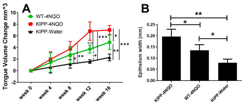
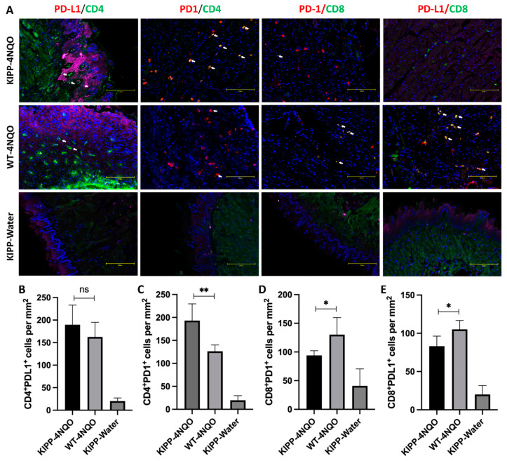
Oral cancer is primarily squamous-cell carcinoma with a 5-year survival rate of approximately 50%. Lysyl oxidase (LOX) participates in collagen and elastin maturation. The propeptide of LOX is released as an 18 kDa protein (LOX-PP) in the extracellular environment by procollagen C-proteinases and has tumor-inhibitory properties. A polymorphism in the propeptide region of LOX (rs1800449, G473A) results in a single amino acid substitution of Gln for Arg. Here we investigated the frequency of rs1800449 in OSCC employing TCGA database resources and determined the kinetics and severity of precancerous oral lesion development in wildtype and corresponding knockin mice after exposure to 4-nitroquinoline oxide (4 NQO) in drinking water. Data show that the OSCC is more common in humans carrying the variant compared to the wildtype. Knockin mice are more susceptible to lesion development. The immunohistochemistry of LOX in mouse tissues and in vitro studies point to a negative feedback pathway of wildtype LOX-PP on LOX expression that is deficient in knockin mice. Data further demonstrate modulations of T cell phenotype in knockin mice toward a more tumor-permissive condition. Data provide initial evidence for rs1800449 as an oral cancer susceptibility biomarker and point to opportunities to better understand the functional mechanism of LOX-PP cancer inhibitory activity.

Keywords: 4 NQO; extracellular matrix; human; lysyl oxidase; mouse model; oral cancer; pathogenesis; polymorphism; tumor suppressor.


Conflict of interest statement

The authors declare no conflict of interest.

Figures

Figure 1
Figure 1
High frequency of rs1800449 (LOX-PP R158Q polymorphism) in OSCC. Chi-square tests: (A) p = 0.0288; n = 156 for cancer patients, and n = 770 for G > A allele frequency (NIH HapMap haplotype map) of the American population. (B) p = 0.0117; n = 156 for cancer patients, and n = 1890 for G > A allele frequency (NIH HapMap haplotype map) of the data set from the human global population. * p < 0.05.
Figure 2
Figure 2
Conserved sequence around human residue R158 in pro-LOX, which when mutated to R158Q compromises LOX-PP tumor-suppressor function. Only the partial C-terminal end of the 14 kDa LOX-PP sequence plus the first few residues of mature LOX domains are shown. The procollagen-C proteinase processing site of pro-LOX that separates LOX-PP from active mature LOX is shown in green. Yellow highlights indicate conserved amino acid sequences.
Figure 3
Figure 3
Experimental design.
Figure 4
Figure 4
Tongue volume measurements in experimental mice. (A) tongue volumes; (B) epithelial thickness. Wildtype mice (WT); knockin mice (KIPP). The error bars indicate +/− SD. Two-way ANOVA, p < 0.05, Tukey’s multiple comparison test, * p < 0.05, ** p < 0.001, *** p < 0.0001 indicate that the mice in the knockin group developed larger volumes of tongues compared to wildtype after 4 NQO treatment.
Figure 5
Figure 5
Hematoxylin and eosin staining of mouse tongues show dysplastic lesions generated in 4 NQO-treated especially in knockin mice with an attenuated response in the one wildtype mouse that developed a papillary lesion. Black arrows show lesion formation. Red arrows show hyperchromatic discohesive cells with increased nuclear to cytoplasmic ratios and white arrows show cohesive epithelial cells. Boxes outline areas shown at higher magnification. Images are representative of 4 mice per group subjected to histology and immunohistochemistry.
Figure 6
Figure 6
Differential PCNA expressions in mouse groups. Knockin mice treated with vehicle exhibit normal basal epithelial expression of PCNA (top panels), while 4 NQO treated mice exhibit lesions with connective-tissue stromal cell expression (middle panels, red arrows), while wildtype mice treated with 4 NQO exhibit suprabasal and basal epithelial staining (bottom panels, blue arrows). The boxes identify regions shown at higher magnification in the right columns. Images are representative of 4 mice per group subjected to histology and immunohistochemistry.
Figure 7
Figure 7
LOX expression is differentially regulated in knockin mice. Samples from knockin mice treated with vehicle show normal low expression of LOX in the basal epithelium (top panel, black arrows), and spinous epithelium. Knockin mice treated with 4 NQO exhibit strong staining in fibroblasts (middle panels, red arrows) and in the suprabasal epithelial cells (middle panel, blue arrows). Wildtype mice express LOX primarily in suprabasal cells (bottom panels, white arrows) with observable staining in basal epithelial cells. The boxes identify regions shown at higher magnification in the right column. Images are representative of 4 mice per group subjected to histology and immunohistochemistry.
Figure 8
Figure 8
G152A polymorphism of LOX-PP attenuated the negative feedback inhibitory effect on LOX expression. Western blot of LY2 cell layers after treatment with 8 µg/mL rArg LOX-PP or rGln-LOXPP.
Figure 9
Figure 9
Differential expression of T cell immune checkpoint PD-L1 and PD-1 in tongue SCC lesions in wildtype (WT) Arg LOX-PP mice and knockin Gln LOX-PP (KIPP) mice. FFPE sections of 4 NQO-induced lesions or controls were stained with PD-L1, PD-1, CD4, and CD8 antibodies, and the images were captured by the EVOS M5000 system. (A) The representative images of CD4+PD-L1+, CD4+PD-1+, CD8+PD-1+, and CD8+PD-L1+ cells in each group. White arrows showed double-positive cells. (BE) The numbers of double-positive cells were quantified by ImageJ and normalized in cells/mm2. Statistical analysis was first calculated by ANOVA and then an unpaired t-test was used to calculate the statistical difference between the Arg LOX-PP and Gln LOX-PP groups. The significance (p-value) is defined as * p < 0.05, ** p < 0.01, and ns p > 0.05. Scale bar, 150 µm.
Figure 10
Figure 10
Summary and hypotheses regarding differences in activity between wildtype Arg LOX-PP and variant Gln LOX-PP in OSCC tissues. While Arg LOX-PP can inhibit LOX production through a negative feedback mechanism variant (left cartoon and Figure 8), Gln LOX-PP loses this inhibitory effect. This ultimately results in more secretion of LOX from oral lesions in ECM and progressing tumor formation (right cartoon). Additional differential effects of Gln LOX-PP occur on T cells in the microenvironment are suggested by high CD4+PD1+ and high CD4+PDL1+ T in the knockin mice (right cartoon and Figure 9), which could lead to enhanced immunosuppression. Created with BioRender.com.

References

    1. Kagan H.M., Trackman P.C. Properties and function of lysyl oxidase. Am. J. Respir. Cell Mol. Biol. 1991;5:206–210. doi: 10.1165/ajrcmb/5.3.206. - DOI - PubMed
    1. Trackman P.C., Bedell-Hogan D., Tang J., Kagan H.M. Post-translational glycosylation and proteolytic processing of a lysyl oxidase precursor. J. Biol. Chem. 1992;267:8666–8671. doi: 10.1016/S0021-9258(18)42494-5. - DOI - PubMed
    1. Barker H.E., Cox T.R., Erler J.T. The rationale for targeting the LOX family in cancer. Nat. Rev. Cancer. 2012;12:540–552. doi: 10.1038/nrc3319. - DOI - PubMed
    1. Palamakumbura A.H., Jeay S., Guo Y., Pischon N., Sommer P., Sonenshein G.E., Trackman P.C. The propeptide domain of lysyl oxidase induces phenotypic reversion of ras-transformed cells. J. Biol. Chem. 2004;279:40593–40600. doi: 10.1074/jbc.M406639200. - DOI - PubMed
    1. Palamakumbura A.H., Vora S.R., Nugent M.A., Kirsch K.H., Sonenshein G.E., Trackman P.C. Lysyl oxidase propeptide inhibits prostate cancer cell growth by mechanisms that target FGF-2-cell binding and signaling. Oncogene. 2009;28:3390–3400. doi: 10.1038/onc.2009.203. - DOI - PMC - PubMed